. 2019 Apr 23;10(2):e00538-19.
doi: 10.1128/mBio.00538-19.

The Structural Basis for a Transition State That Regulates Pore Formation in a Bacterial Toxin

Affiliations

The Structural Basis for a Transition State That Regulates Pore Formation in a Bacterial Toxin

Kristin R Wade et al. mBio. .

Abstract

The cholesterol-dependent cytolysin (CDC) genes are present in bacterial species that span terrestrial, vertebrate, and invertebrate niches, which suggests that they have evolved to function under widely different environmental conditions. Using a combination of biophysical and crystallographic approaches, we reveal that the relative stability of an intramolecular interface in the archetype CDC perfringolysin O (PFO) plays a central role in regulating its pore-forming properties. The disruption of this interface allows the formation of the membrane spanning β-barrel pore in all CDCs. We show here that the relative strength of the stabilizing forces at this interface directly impacts the energy barrier posed by the transition state for pore formation, as reflected in the Arrhenius activation energy (Ea) for pore formation. This change directly impacts the kinetics and temperature dependence of pore formation. We further show that the interface structure in a CDC from a terrestrial species enables it to function efficiently across a wide range of temperatures by minimizing changes in the strength of the transition state barrier to pore formation. These studies establish a paradigm that CDCs, and possibly other β-barrel pore-forming proteins/toxins, can evolve significantly different pore-forming properties by altering the stability of this transitional interface, which impacts the kinetic parameters and temperature dependence of pore formation.IMPORTANCE The cholesterol-dependent cytolysins (CDCs) are the archetype for the superfamily of oligomeric pore-forming proteins that includes the membrane attack complex/perforin (MACPF) family of immune defense proteins and the stonefish venom toxins (SNTX). The CDC/MACPF/SNTX family exhibits a common protein fold, which forms a membrane-spanning β-barrel pore. We show that changing the relative stability of an extensive intramolecular interface within this fold, which is necessarily disrupted to form the large β-barrel pore, dramatically alters the kinetic and temperature-dependent properties of CDC pore formation. These studies show that the CDCs and other members of the CDC/MACPF/SNTX superfamily have the capacity to significantly alter their pore-forming properties to function under widely different environmental conditions encountered by these species.

Keywords: cold denaturation; complement; gasdermin; perforin; water network.


Figures

FIG 1
FIG 1
The CDC mechanism. (A) Top-down view of a model of the PFO oligomeric pore complex (monomers shown in alternating red and blue) based on the 4.5-Å resolution cryo-electron microscopy structure of the related CDC, PLY (40). (B) Cutaway view of the pore viewed from inside the lumen. (C) The structure of soluble PFO (22) showing the location of the α-helical bundles (αHBs, green) and the location of N197 (purple space-filled atoms) at the interface between domain 3 and domains 1 and 2 (D3-D1,2). Within the prepore structure, this interface is necessarily disrupted to refold the twin αHBs into the extended transmembrane β-hairpins shown in the structure (right panel) of a PFO monomer extracted from the oligomeric complex, showing that N197 faces the luminal side of the β-barrel in the transmembrane β-hairpin 1 (17, 18). Shown in red in panels C and D is D2, which acts as a ratchet to lower domains D1 and D3 ∼40 Å nearer the membrane so that the extended transmembrane β-hairpins can cross the bilayer to form the β-barrel pore (40–42). (D) Domains D4 of the structures in panel C were overlaid to illustrate the vertical collapse of domains D1 to D3 upon pore formation as viewed from the pore lumen in panel B. Structures were generated using UCSF Chimera (43).
FIG 2
FIG 2
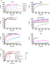
Pore formation rates of CDCs at various temperatures. (A) The rate of pore formation for the indicated CDCs from 9 to 37°C was measured by the release of the fluorescence marker carboxyfluorescein (CF) over time from cholesterol-rich liposomes. Each assay was normalized to the maximum emission obtained with PFON197W at 37°C. (B) The 30.2°C and 37°C data for PFO and PFOV97G-S98A are overlaid to show that at permissive temperatures the latter exhibits a higher rate of pore formation determined by the time to 50% marker release (t1/2). Each toxin (36 nM final concentration) was injected into HBS buffer (2 ml) containing carboxyfluorescein (CF)-loaded liposomes (20 μl) at 30 s, and pore formation was monitored by the increase in fluorescence intensity of the CF, as its fluorescence emission was dequenched by release from the liposomes upon pore formation.
FIG 3
FIG 3
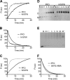
Structural transitions required for oligomerization and pore formation are accelerated in PFON197W. (A) The rate of membrane binding was determined by following the increase in the emission over time of the UDP tryptophan residues in D4 (19), as monomers of PFO (solid line) and PFON197W (dashed line) bind to the liposomes. (B) The rate of monomer oligomerization into the prepore complex was determined by following the disengagement of β5 from β4. This disengagement was detected by monitoring the decrease in the emission over time of NBD-modified cysteine-substituted V322 in PFOV322C (solid line) or PFOV322C-N197W (dashed line). V322 is buried beneath the α1β5 loop (Fig. 4A, left panel), and upon the disengagement of β5 from β4 during oligomerization, NBD is exposed to water, which quenches its emission (20). (C) The membrane insertion of the β-barrel was determined by the increase in the fluorescence emission of NBD positioned on cysteine-substituted A215 (18), a membrane-facing residue in transmembrane β-hairpin 1, in PFOA215C (solid line) and PFOA215C-N197W (dashed line). All data were normalized to 100 and are representative of 3 independent experiments. (D and E) PFO, PFON197W, or DLY was injected into buffer containing cholesterol-rich liposomes, and samples were withdrawn at the times indicated and immediately placed into SDS-PAGE sample buffer to quench the assembly of the pore. The samples were then separated by SDS-AGE to separate the monomer (M) from the oligomer (O). Note that the disappearance of the DLY monomer is rapid, as it assembles into the pore complex, which tends to form a smear in the absence of heat. *, same as the DLY 30-min sample that was additionally heated to 95°C for 3 min to show that the smeared oligomers migrate as a single band when heat is applied. (F) The rate of the disengagement of β5 from β4 in PFOS97G-V98A compared to PFO was measured as described in the legend to panel B. All fluorescence assays were carried out at 37°C. M, soluble toxin monomer in the absence of liposomes.
FIG 4
FIG 4
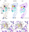
The solution of the crystal structures of DLY and PFON197W reveals differences with PFO at the D3-D1,2 interface. (A) The α-carbon backbone representation of the crystal structures of the previously solved structure of PFO (22) and the structures solved here for PFON197W and DLY. The waters at the D3-D1,2 interface are shown for all three proteins as light blue transparent Van der Waals (VDW) representations. Also shown are the conserved diglycine pairs in D3 (yellow space-filled atoms), the S97 and V98 pair (red and blue space-filled atoms, respectively), Y358 (gold space-filled atoms), the α-helical bundles (αHBs) that ultimately refold to form transmembrane β-hairpins 1 and 2 (cyan), the α1β5 loop (dark red), and the structures of the membrane binding interface: the cholesterol recognition/binding threonine-leucine pair (CRM) and the undecapeptide (UDP, shown in black). (B) Stereo image showing the region boxed in PFO (tan) in panel A overlaid with the same regions in PFON197W (light blue) and DLY (purple), showing the displacement of the D96-Y103 loop (shown by the arrows) in PFON197W and specifically showing the displacement of S97 and V98 from their interaction with the conserved glycine pair (G324, G325), which serves as a flexible linker to allow the rotation of β5 away from β4 (20) and from Y358. The positions of the analogous residues (A67 and Y68) are shown for DLY, and the figure shows that the analogous residue for Y358 in PFO is an alanine in DLY.
FIG 5
FIG 5
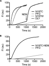
Pore formation by PFON197C at low temperature modified with NEM before and after prepore assembly. (A) Pore formation was monitored over time by CF release from liposomes treated with PFO (±NEM), PFON197W, PFON197C (±NEM), and DLY at 6°C. CF release by PFO and PFON197C was monitored for 1,000 s to allow for prepore formation, and then N-ethylmaleimide (NEM) was injected (final concentration of 100 μM) to modify the sulfhydryl of N197C. As shown, NEM triggers the rapid conversion of prepore PFON197C but has no effect on the low rate of pore formation by PFO. (B) The cysteine sulfhydryl of PFON197C was labeled with NEM prior to injecting it into liposomes at 6°C compared to PFO. Note that the rate of pore formation by PFON197C is higher when the prepore is allowed to first assemble followed by the injection of NEM in panel A than when PFON197C is labeled with NEM prior to its injection into the liposomes in panel B.
FIG 6
FIG 6
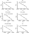
Arrhenius activation energies of pore formation by PFO, DLY, and their derivatives. The Arrhenius activation energies (Ea) were determined for the proteins shown. The initial velocities (k0) of pore formation (the linear portion of the marker release curve, as in Fig. 2) at the various temperatures were used to determine the Ea. Each analysis was carried out in quadruplicate, and the standard error (SE) for 1/k0 at each temperature is shown. Those SEs with error bars smaller than the symbols are not visible.
FIG 7
FIG 7
DLY D3-D1,2 interface near L162. Leucine 162 of DLY is located in a hydrophobic pocket primarily composed of aromatic residues F25, Y56, F60, and A58, whereas the analogous residues in PFO are primarily hydrophilic residues. Also shown is the location of N197 in PFO, which has been replaced by G165 in DLY. The interaction energies (IE) (http://bioinfo.uochb.cas.cz/INTAA/energy/) of the major residues that interact with DLY L162 and its analog in PFO, S194, are shown for each protein.

References

    1. Tweten RK, Hotze EM, Wade KR. 2015. The unique molecular choreography of giant pore formation by the cholesterol-dependent cytolysins of gram-positive bacteria. Annu Rev Microbiol 69:323–340. doi:10.1146/annurev-micro-091014-104233. - DOI - PMC - PubMed
    1. Dunstone MA, Tweten RK. 2012. Packing a punch: the mechanism of pore formation by cholesterol dependent cytolysins and membrane attack complex/perforin-like proteins. Curr Opin Struct Biol 22:342–349. doi:10.1016/j.sbi.2012.04.008. - DOI - PMC - PubMed
    1. Ellisdon AM, Reboul CF, Panjikar S, Huynh K, Oellig CA, Winter KL, Dunstone MA, Hodgson WC, Seymour J, Dearden PK, Tweten RK, Whisstock JC, McGowan S. 2015. Stonefish toxin defines an ancient branch of the perforin-like superfamily. Proc Natl Acad Sci U S A 112:15360–15365. doi:10.1073/pnas.1507622112. - DOI - PMC - PubMed
    1. Hotze EM, Le HM, Sieber JR, Bruxvoort C, McInerney MJ, Tweten RK. 2013. Identification and characterization of the first cholesterol-dependent cytolysins from Gram-negative bacteria. Infect Immun 81:216–225. doi:10.1128/IAI.00927-12. - DOI - PMC - PubMed
    1. Grady EN, MacDonald J, Liu L, Richman A, Yuan ZC. 2016. Current knowledge and perspectives of Paenibacillus: a review. Microb Cell Fact 15:203. doi:10.1186/s12934-016-0603-7. - DOI - PMC - PubMed

Publication types

MeSH terms

Substances

LinkOut - more resources