. 2019 Apr 23;10(2):e02904-18.
doi: 10.1128/mBio.02904-18.

Stable and Highly Immunogenic MicroRNA-Targeted Single-Dose Live Attenuated Vaccine Candidate against Tick-Borne Encephalitis Constructed Using Genetic Backbone of Langat Virus

Affiliations

Stable and Highly Immunogenic MicroRNA-Targeted Single-Dose Live Attenuated Vaccine Candidate against Tick-Borne Encephalitis Constructed Using Genetic Backbone of Langat Virus

Konstantin A Tsetsarkin et al. mBio. .

Abstract

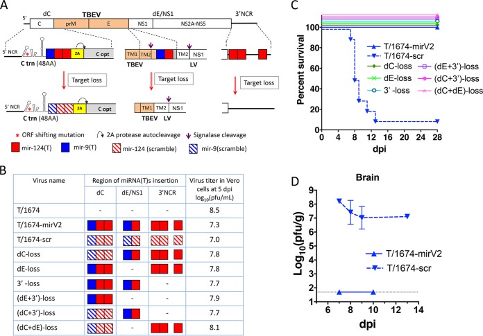
Tick-borne encephalitis virus (TBEV), a member of the genus Flavivirus, is one of the most medically important tick-borne pathogens of the Old World. Despite decades of active research, attempts to develop of a live attenuated virus (LAV) vaccine against TBEV with acceptable safety and immunogenicity characteristics have not been successful. To overcome this impasse, we generated a chimeric TBEV that was highly immunogenic in nonhuman primates (NHPs). The chimeric virus contains the prM/E genes of TBEV, which are expressed in the genetic background of an antigenically closely related, but less pathogenic member of the TBEV complex-Langat virus (LGTV), strain T-1674. The neurovirulence of this chimeric virus was subsequently controlled by robust targeting of the viral genome with multiple copies of central nervous system-enriched microRNAs (miRNAs). This miRNA-targeted T/1674-mirV2 virus was highly stable in Vero cells and was not pathogenic in various mouse models of infection or in NHPs. Importantly, in NHPs, a single dose of the T/1674-mirV2 virus induced TBEV-specific neutralizing antibody (NA) levels comparable to those seen with a three-dose regimen of an inactivated TBEV vaccine, currently available in Europe. Moreover, our vaccine candidate provided complete protection against a stringent wild-type TBEV challenge in mice and against challenge with a parental (not miRNA-targeted) chimeric TBEV/LGTV in NHPs. Thus, this highly attenuated and immunogenic T/1674-mirV2 virus is a promising LAV vaccine candidate against TBEV and warrants further preclinical evaluation of its neurovirulence in NHPs prior to entering clinical trials in humans.IMPORTANCE Tick-borne encephalitis virus (TBEV) is one of the most medically important tick-borne pathogens of the Old World. Despite decades of active research, efforts to develop of TBEV live attenuated virus (LAV) vaccines with acceptable safety and immunogenicity characteristics have not been successful. Here we report the development and evaluation of a highly attenuated and immunogenic microRNA-targeted TBEV LAV.

Keywords: Langat virus; chimeric virus; live attenuated virus vaccine; microRNA; tick-borne encephalitis virus.


Figures

FIG 1
FIG 1
Construction of the chimeric miRNA-targeted TBEV/LGTVs and their immunogenicity in NHPs. (A) Schematic representation of the chimeric T/1674 virus. The white boxes correspond to LGTV strain 1674 sequences. Orange boxes depict TBEV sequences (strain Sofjin). The genetic organization of the miRNA-targeting cassettes that were used to generate T/1674-mirV2 and positions of their insertion into the dC, dE/NS1, and 3′NCR regions of the T/1674 virus is indicated. C trn (48AA), replication promoter region of the C gene of LGTV; TM1 and TM2, transmembrane helical regions of E protein. Gray boxes represent the codon-optimized sequences of the C gene of TBEV and TM regions of LGTV. (B) Characterization of chimeric TBEV/LGTVs that were used in the NHP immunogenicity study. Compositions of the miRNA-targeting cassettes inserted into the dC, dE/NS1 and 3′NCR regions of T/1674 virus are depicted. Red boxes indicate the targets for mir-124; blue boxes indicate the targets for mir-9. Viruses were purified by the one-step terminal dilution method. Working stocks were generated after the second consecutive passage in Vero cells followed by virus titration in Vero cells. Stability of miRNA(T)s was assessed after 10 passages in Vero cells by sequencing analysis. The crosses (+) indicate that all miRNA targets in the virus remained stable at the passage 10. (C and D) Experimental design (C) of the study performed to compare viremia, immunogenicity, and protective efficacy data for the chimeric TBEV/LGTVs in NHPs (D). Rhesus macaques in groups of three or four were subjected to mock inoculation or infected subcutaneously with 105 PFU of miRNA-targeted viruses. Monkeys were bled on dpi 1 to 7 to determine the duration of viremia (expressed as mean number of viremia days per animal in the group) and the mean peak viral titer in the serum of each animal in the group [expressed as log10(PFU/ml)]. On dpi 28 and 56, animals were bled to determine NA titer (expressed as geometric mean [GMT] for the group) using the 50% plaque reduction neutralization (PRNT50) assay. At dpi 29, animals were challenged s.c. with 105 PFU of T/E5 or T/1674 virus, followed by evaluation of viremia duration and peak virus titer in serum. Virus titer in serum was determined by focus-forming assay in LLC-MK2 cells, and data are expressed as log10(PFU/ml). NT, not tested. Serum samples from four animals s.c. inoculated with a formalin-inactivated TBEV vaccine “Encepur” in three human doses (3 × 0.5 ml) on days 0, 7, and 21 and collected for neutralization assay 21 days after the third dose were from our previous studies (13).
FIG 2
FIG 2
Immunogenicity, protective efficacy, and replication of the miRNA-targeted TBEV/LGTVs in C3H mice. (A to C) C3H mice (n = 5) were inoculated with T/1674, T/1674-mirV1, or T/1674-mirV2 virus or with diluent (mock). At day 29 postimmunization, animals were challenged i.p. with 105 PFU of T/1674 (n = 5) and monitored for neurological signs. (A) Survival of C3H mice (n = 5) after i.p. inoculation with 105 PFU of T/1674, T/1674-mirV1, and T/1674-mirV2 viruses. (B) Neutralizing antibody (NA) titer in the serum of C3H mice on day 28 postimmunization with miRNA-targeted viruses and on day 27 postchallenge with T/1674. The NA titer was determined using the 50% plaque reduction neutralization (PRNT50) assay against the T/E5 virus. Differences in NA titer in the mouse sera were compared using the Mann-Whitney test (ns, not statistically significant [P > 0.05]). (C) Survival of immunized C3H mice (n = 5) after the challenge with 105 PFU T/1674 virus. (D) C3H mice (n = 10) were inoculated i.p. with 105 PFU of T/1674-mirV1 or T/1674-mirV2 or with diluent (mock). At day 29 postimmunization, animals were challenged i.p. with 105 PFU of a wild-type TBEV (strain Hypr). The graph shows the survival of immunized mice. (E to K) Kinetics of T/1674 and T/1674-mirV2 virus replication in the brain (E), serum (F), kidney (G), lung (H), spleen (I), muscle (J), and pancreas (K) of C3H mice. Three-week-old mice were infected i.p. with 105 PFU of T/1674 or T/1674-mirV2 virus. At 1, 3, 5, 7, and 10 dpi, mice were sacrificed (3 animals per time point in each group) and virus titers in the organs and serum were determined by titration in Vero cells. The dashed lines indicate the limits of virus detection: 1.5 log10(PFU/ml) for serum; 1.7 log10(PFU/g) for brain (E), kidney (G), lung (H), spleen (I), and muscle (J); and 2.7 log10(PFU/g) for pancreas (K). Differences between T/1674 and T/1674-mirV2 titers in mouse serum or organs were compared using two-way ANOVA.
FIG 3
FIG 3
Pathogenesis of the miRNA-targeted TBEV/LGTV in the brain of newborn mice. (A) Survival of newborn SW mice (n = 10 per group) after i.c. infection with 103 PFU of T/1674-mirV1 and T/1674-mirV2 viruses or i.c. infection with 10−1, 10°, or 103 PFU of T/1674 virus. (B) Growth kinetics of parental (T/1674) and miRNA-targeted viruses in the brain of newborn mice after i.c. infection with a dose of 103 PFU. For each time point, brains from three pups per virus were collected to determine virus loads by titration in Vero cells. Mean virus titers ± standard deviations (SD) of brain homogenates are shown. Differences in growth kinetics between viruses were compared using two-way ANOVA. The dashed line indicates the limit of virus detection (1.7 log10 PFU/g of brain tissue). A crosshatch symbol (#) indicates that after the indicated time point, collection of brain samples was terminated due to the death of the T/1674-infected animals. (C to K) Immunohistochemical analysis. Representative areas of the cerebral cortex of suckling mice were inoculated i.c. with a dose consisting of 103 PFU of the T/1674 virus (4 dpi) or the T/1674-mirV2 virus (22 dpi) or were subjected to mock inoculation (22 dpi). (C to E) Immunoreactivity (IR) for TBEV antigens. (F to H) Iba1-IR images showing the morphology of microglia. (I to K) Changes in the integrity of somatodendritic neuronal compartments revealed by MAP-2 IR. Bars, 50 μm.
FIG 4
FIG 4
Phenotypic and genetic stability of the T/1674-mirV2 virus during prolonged replication in SCID mice. (A) Experimental design. Three-week-old SCID mice (males; n = 5 per group) were infected i.p. with 105 PFU of T/1674 or T/1674-mirV2 virus and monitored for onset of neurological disease for 53 dpi. At indicated intervals postinfection, mice were bled to determine virus titers in the serum. (B) Survival of SCID mice infected with T/1674 or T/1674-mirV2. Differences in the survival rates of paired groups of mice were compared using the log rank test. (C) Mean virus titer ± SD in the serum of mice. A crosshatch symbol (#) indicates that serum samples from the T/1674-infected mice were not collected after dpi 7 due to the death of the animals. (D) Comparison of the T/1674 and T/1674-mirV2 virus titers in brain and spleen of SCID mice. Brains and spleens were collected from T/1674-infected mice at the time of death (8 to 11 dpi) and from T/1674-mirV2-infected mice at 53 dpi. Differences between viral titers were compared using the unpaired two-tailed t test (****, P< 0.0001; ns, not significant [P > 0.05]). (E and F) Stability of miRNA(T)s with respect to the T/1674-mirV2 virus isolated from the brain (E) and serum (F) of SCID mice at 53 dpi. Regions containing miRNA-targeting cassettes (dC, E/NS1, and 3′NCR) were amplified and sequenced. Solid-colored boxes represent target sequences for mir-124 (red) and mir-9 (blue) in the T/1674-mirV2 genome that remained stable at 53 dpi. The striped blue box indicates the mir-9(T) sequence, which acquired heterogenicity in nt 8 of the target (as shown in the electropherograms below the panels).
FIG 5
FIG 5
Resistance of the T/1674-mirV2 virus to mutational miRNA(T) instability. (A) Diagram depicting genetic modifications of the miRNA-targeting cassettes located in the dC and dE/NS1 regions and in the 3′NCR of the T/1674-mirV2 virus. These modifications were used to construct viruses containing reduced numbers of miRNA-targeting cassettes. (B) Schematic representation of the viruses containing reduced numbers (compared to T/1674-mirV2) of miRNA-targeting cassettes in their genomes and titers of these viruses in Vero cell supernatants at 5 dpi. Striped red and blue boxes indicate that corresponding mir-124(T) and mir-9(T) sequences contain multiple nucleotide substitutions. Dashes (–) indicate that the corresponding miRNA-targeting region of the T/1674-mirV2 virus was replaced (deleted) with the sequence of a parental T/1674 virus. (C) Survival of C3H mice (n = 10) after i.p. infection with 105 PFU of the viruses depicted in panel B. (D) The titer of T/1674-scr virus in the brain of morbid C3H mice. Brains were collected from mice that had succumbed to neurologic disease in the experiment depicted in panel C, and virus titers were determined by titration in Vero cells. The titers of T/1674-mirV2 virus in the brain of healthy C3H mice at 7 and 10 dpi are given in Fig. 2E and are presented here for comparison.

Similar articles

Cited by

References

    1. Lehrer AT, Holbrook MR. 2011. Tick-borne encephalitis vaccines. J Bioterror Biodef 2011:3. doi:10.4172/2157-2526.S1-003. - DOI - PMC - PubMed
    1. Ishikawa T, Yamanaka A, Konishi E. 2014. A review of successful flavivirus vaccines and the problems with those flaviviruses for which vaccines are not yet available. Vaccine 32:1326–1337. doi:10.1016/j.vaccine.2014.01.040. - DOI - PubMed
    1. Pletnev AG, Men R. 1998. Attenuation of the Langat tick-borne flavivirus by chimerization with mosquito-borne flavivirus dengue type 4. Proc Natl Acad Sci U S A 95:1746–1751. doi:10.1073/pnas.95.4.1746. - DOI - PMC - PubMed
    1. Thind IS, Price WH. 1966. A chick embryo attenuated strain (TP21 E5) of Langat virus. I. Virulence of the virus for mice and monkeys. Am J Epidemiol 84:193–213. doi:10.1093/oxfordjournals.aje.a120633. - DOI - PubMed
    1. Smith CE. 1956. A virus resembling Russian spring-summer encephalitis virus from an ixodid tick in Malaya. Nature 178:581–582. doi:10.1038/178581a0. - DOI - PubMed

Publication types

MeSH terms

LinkOut - more resources