. 2019 Jun 26;39(26):5222-5233.
doi: 10.1523/JNEUROSCI.2204-18.2019. Epub 2019 Apr 23.

Transcriptional Regulation of Voltage-Gated Sodium Channels Contributes to GM-CSF-Induced Pain

Affiliations

Transcriptional Regulation of Voltage-Gated Sodium Channels Contributes to GM-CSF-Induced Pain

Fan Zhang et al. J Neurosci. .

Abstract

Granulocyte-macrophage colony-stimulating factor (GM-CSF) induces the production of granulocyte and macrophage populations from the hematopoietic progenitor cells; it is one of the most common growth factors in the blood. GM-CSF is also involved in bone cancer pain development by regulating tumor-nerve interactions, remodeling of peripheral nerves, and sensitization of damage-sensing (nociceptive) nerves. However, the precise mechanism for GM-CSF-dependent pain is unclear. In this study, we found that GM-CSF is highly expressed in human malignant osteosarcoma. Female Sprague Dawley rats implanted with bone cancer cells develop mechanical and thermal hyperalgesia, but antagonizing GM-CSF in these animals significantly reduced such hypersensitivity. The voltage-gated Na+ channels Nav1.7, Nav1.8, and Nav1.9 were found to be selectively upregulated in rat DRG neurons treated with GM-CSF, which resulted in enhanced excitability. GM-CSF activated the Janus kinase 2 (Jak2)-signal transducer and activator of transcription protein 3 (Stat3) signaling pathway, which promoted the transcription of Nav1.7-1.9 in DRG neurons. Accordingly, targeted knocking down of either Nav1.7-1.9 or Jak2/Stat3 in DRG neurons in vivo alleviated the hyperalgesia in male Sprague Dawley rats. Our findings describe a novel bone cancer pain mechanism and provide a new insight into the physiological and pathological functions of GM-CSF.SIGNIFICANCE STATEMENT It has been reported that granulocyte-macrophage colony-stimulating factor (GM-CSF) plays a key role in bone cancer pain, yet the underlying mechanisms involved in the GM-CSF-mediated signaling pathway in nociceptors is not fully understood. Here, we showed that GM-CSF promotes bone cancer-associated pain by enhancing the excitability of DRG neurons via the Janus kinase 2 (Jak2)-signal transducer and activator of transcription protein 3 (Stat3)-mediated upregulation of expression of nociceptor-specific voltage-gated sodium channels. Our study provides a detailed understanding of the roles that sodium channels and the Jak2/Stat3 pathway play in the GM-CSF-mediated bone cancer pain; our data also highlight the therapeutic potential of targeting GM-CSF.

Keywords: DRG; GM-CSF; Jak2-Stat3; bone cancer pain; neural excitability; voltage-gated sodium channels.


Figures

Figure 1.
Figure 1.
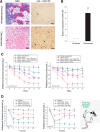
Role of GM-CSF in bone cancer metastases pain. A, High expression level of GM-CSF in osteosarcoma tissue sample. H&E staining of chondroma and osteosarcoma is shown on the left; immunohistochemical staining for GM-CSF, indicated by arrows in chondroma and osteosarcoma, is shown on the right. B, Summary results for immunohistochemical staining for GM-CSF (n = 8/group; unpaired t test: t = 5.69, *p = 0.0013). C, Effect of antibodies against GM-CSF or GM-CSFR (10 μg) and GM-CSF analog E21R (a competitive antagonist of GM-CSF, 25 μg/μl, 3 μl) on mechanical (left) and thermal (right) nociceptive responses in a bone cancer pain model in rats. The mechanical paw withdrawal threshold and thermal paw withdrawal latency were measured at 3, 7, 11, 14, 17, and 21 d for the control group (black line + squares), the bone cancer group (red line + circles), the bone cancer + antibody against GM-CSF group (blue line + triangles), the bone cancer + antibody against GM-CSFR group (pink line + triangles), and the bone cancer + E21R group (green line + triangles). Two-way ANOVA followed by Bonferroni post hoc tests revealed a significant effect of treatment (F(4,150) = 29.49, p = 0) and time (F(5,150) = 5.06, p < 0.0001), but no interaction between the two (F(20,150) = 0.71, p = 0.81; left); and a significant effect of treatment (F(4,240) = 15.10, p < 0.0001), time (F(4,240) = 2.56, p = 0.028), and an interaction between the two (F(20,240) = 2.20, p = 0.003; right). *p < 0.05 compared with sham group; #p < 0.05 with respect to the corresponding bone cancer group. (D) Dose-dependent effects of GM-CSF on paw withdrawal threshold to mechanical stimulus (left) and on paw withdrawal latency to noxious heat (right) at 1, 2, 5, 12, and 24 h following focal DRG application via DRG cannula. Number of experiments is indicated as n in each panel. Two-way ANOVA followed by Bonferroni post hoc tests revealed a significant effect of dose (F(3,345) = 29.30, p < 0.0001) but not of time (F(3,345) = 0.15, p = 0.96) or interaction between the two (F(12,345) = 0.37, p = 0.97; left). Right, A significant effect of dose (F(3,350) = 70.95, p = 0) and an interaction between dose and time (F(12,350) = 3.71, p < 0.0001), but the effect of time did not reach significance (F(3,350) = 2.09, p = 0.08). *p < 0.05 compared with the vehicle saline.
Figure 2.
Figure 2.
Effect of GM-CSF on the excitability of small-sized DRG neurons. A, Representatives of action potentials evoked by depolarizing current pulse (left), recorded from small-sized DRG neurons. B, Summary results for the effect of GM-CSF on numbers of action potential induced by increasing amplitudes of depolarizing currents. Two-way ANOVA followed by Bonferroni post hoc tests revealed a significant effect of treatment (F(1,1057) = 22.45, p < 0.0001) and injected currents (F(9,1057) = 7.82, p < 0.0001), but not of a significant interaction between the two (F(9,1057) = 0.43, p = 0.92). *p < 0.05 compared with the control. C, Single action potentials from A with expanded time scales. D, Summary results for the effect of GM-CSF on the threshold potential, rheobase current, and RMP (unpaired t test, *p < 0.05 compared with the control).
Figure 3.
Figure 3.
Effect of GM-CSF on the current amplitude and expression level of Nav1.7, Nav1.8, and Nav1.9 channels. A, Relative mRNA expression of Nav1.7, Nav1.8, Nav1.9, Kv4.2, TMEM16A, P2X3, KCNQ2, and KCNQ3 in cultured DRG cells after incubation of GM-CSF (200 ng/ml) at 24 h (n = 9, unpaired t test, *p < 0.05 compared with the control). B, Relative mRNA expression of Nav1.7, Nav1.8, and Nav1.9 in DRG neurons of bone cancer pain at the seventh day. (n = 6, unpaired t test, *p < 0.05 compared with the control). C, Typical current traces and current density–voltage relationship of total TTX-S, TTX-R, Nav1.8, and Nav1.9 Na+ currents in cultured DRG cells after incubation with GM-CSF (200 ng/ml) for 24 h. D, Western blot analysis of expression levels of Nav1.7, Nav1.8, and Nav1.9 proteins in DRG neurons treated with GM-CSF (200 ng/ml) for 18 h (n = 3, unpaired t test, *p < 0.05 compared with the control).
Figure 4.
Figure 4.
Downregulation of Nav1.7, Nav1.8, and Nav1.9 reverses nociceptive behavior evoked by GM-CSF. AC, Application of AS ODNs (ASOs) in DRGs against Nav1.7, Nav1.8, and Nav1.9 (each ASO, 12.5 μg/rat, 5 μl) significantly reduced the mRNA expression level of Nav1.7, Nav1.8, and Nav1.9 increased by GM-CSF treatment (A), and alleviated mechanical (B) and thermal hyperalgesia (C) produced by the focal GM-CSF (200 ng) application. For A: n = 6; unpaired t test, *p < 0.05 compared with control; #p < 0.05 with respect to the corresponding GM-CSF. For B, Left, two-way ANOVA followed by Bonferroni post hoc tests revealed a significant effect of treatment (F(3,305) = 109.59, p = 0), but not time (F(4,305) = 0.78, p = 0.54) or interaction between the two (F(12,305) = 0.65, p = 0.80). Middle, There was a significant effect of treatment (F(3,305) = 80.53, p = 0), but not time (F(4,305) = 0.20, p = 0.94) or interaction between the two (F(12,305) = 0.42, p = 0.95). Right, There was a significant effect of treatment (F(3,335) = 109.87, p = 0), but not time (F(4,335) = 0.89, p = 0.47) or interaction between the two (F(12,335) = 0.37, p = 0.97). For C, Left, Two-way ANOVA followed by Bonferroni post hoc tests revealed a significant effect of treatment (F(3,295) = 168.25, p = 0) and interaction between treatment and time (F(12,295) = 3.73, p < 0.0001), but the effect of time was not significant (F(4,295) = 1.34, p = 0.25). Middle, There was a significant effect of treatment (F(3,295) = 336.23, p = 0) and an interaction between treatment and time (F(12,295) = 2.04, p = 0.02), but the effect of time was not significant (F(4,295) = 1.05, p = 0.38). Right, There was a significant effect of treatment (F(3,250) = 274.66, p = 0) and interaction between treatment and time (F(12,335) = 5.38, p < 0.0001), but the effect of time was not significant (F(4,335) = 0.71, p = 0.74). *p < 0.05 compared with the vehicle saline; n = 6, #p < 0.05 with respect to the corresponding GM-CSF.
Figure 5.
Figure 5.
GM-CSF increases the mRNA expression level of Nav1.7, Nav1.8, and Nav1.9 channels through the Jak2–Stat3 signaling pathway. A, Relative expression of p-Jak1, p-Jak2, p-Jak3, p-stat3, and p-stat5 in DRG neurons after incubation with GM-CSF for 25 min (n = 3; unpaired t test, *p < 0.05 compared with control). B, Relative mRNA expression level of Nav1.7, Nav1.8, and Nav1.9 in DRG neurons incubated with GM-CSF in the absence or presence of AG-490 (10 μm) and stattic (20 μm) for 4 h (n = 4–6; unpaired t test, *p < 0.05 compared with control; #p < 0.05 with respect to the corresponding GM-CSF). C, Relative Luciferase activity in HEK 293 cells transfected with reporter vector containing Nav1.7, Nav1.8, and Nav1.9 promoter regions (pGL3) coexpressed with either pcDNA3.1 (control) or pcDNA3.1-Stat3 cDNA (n = 3; unpaired t test, *p < 0.05 compared with control). D–F, Relative mRNA level of Nav1.7, Nav1.8, and Nav1.9 in ipsilateral DRGs (L5) of rats receiving AS ODNs (ASOs) against different Jak and Stat signaling molecules (12.5 mg/rat, 5 μl; n = 6, unpaired t test, *p < 0.05 compared with control; #p < 0.05 with respect to the corresponding GM-CSF). G–I, Effect of ASOs against Jak and Stat signaling molecules (12.5 mg/rat, 5 μl) on hyperalgesia responses to mechanical and thermal stimuli induced by GM-CSF. ASOs were given through the DRG cannula for 4 d, and then GM-CSF (200 ng) was given. For G, Left, Two-way ANOVA followed by Bonferroni post hoc tests revealed a significant effect of treatment (F(3,415) = 125.38, p = 0), but not time (F(4,415) = 0.54, p = 0.70) or interaction between the two (F(12,415) = 0.73, p = 0.73). Right, There was a significant effect of treatment (F(3,425) = 77.18, p = 0), but not time (F(4,425) = 1.24, p = 0.29) or interaction between the two (F(12,425) = 1.65, p = 0.07). For H, Left, Two-way ANOVA followed by Bonferroni post hoc tests revealed a significant effect of treatment (F(3,415) = 110.97, p = 0), but not time (F(4,415) = 0.38, p = 0.82) or interaction between the two (F(12,415) = 0.43, p = 0.79). Right, There was a significant effect of treatment (F(3,440) = 115.88, p = 0), but not time (F(4,440) = 1.40, p = 0.25) or interaction between the two (F(12,440) = 1.13, p = 0.33). For I, Left, Two-way ANOVA followed by Bonferroni post hoc tests revealed a significant effect of treatment (F(3,345) = 143.47, p = 0), but not time (F(4,415) = 0.74, p = 0.57) or interaction between the two (F(12,415) = 0.45, p = 0.94). Right, There was a significant effect of treatment (F(3,440) = 111.32, p = 0), but not time (F(4,440) = 0.88, p = 0.47) or an interaction between the two (F(12,440) = 0.94, p = 0.50). n = 6–8; *p < 0.05 compared with the vehicle saline; #p < 0.05 with respect to the corresponding GM-CSF.

Similar articles

Cited by

References

    1. Alexander WS. (2016) In vivo at last: demonstrating the biological credentials and clinical potential of GM-CSF. Exp Hematol 44:669–673. 10.1016/j.exphem.2016.01.006 - DOI - PubMed
    1. Bali KK, Venkataramani V, Satagopam VP, Gupta P, Schneider R, Kuner R (2013) Transcriptional mechanisms underlying sensitization of peripheral sensory neurons by granulocyte-/granulocyte-macrophage colony stimulating factors. Mol Pain 9:48. 10.1186/1744-8069-9-48 - DOI - PMC - PubMed
    1. Blanchard MG, Rash LD, Kellenberger S (2012) Inhibition of voltage-gated Na(+) currents in sensory neurones by the sea anemone toxin APETx2. Br J Pharmacol 165:2167–2177. 10.1111/j.1476-5381.2011.01674.x - DOI - PMC - PubMed
    1. Chaplan SR, Bach FW, Pogrel JW, Chung JM, Yaksh TL (1994) Quantitative assessment of tactile allodynia in the rat paw. J Neurosci Methods 53:55–63. - PubMed
    1. Cheng X, Dib-Hajj SD, Tyrrell L, Te Morsche RH, Drenth JP, Waxman SG (2011) Deletion mutation of sodium channel Na(V)1.7 in inherited erythromelalgia: enhanced slow inactivation modulates dorsal root ganglion neuron hyperexcitability. Brain 134:1972–1986. 10.1093/brain/awr143 - DOI - PubMed

Publication types

MeSH terms

Substances