. 2019 May 21;39(5):BSR20182051.
doi: 10.1042/BSR20182051. Print 2019 May 31.

In vitro differentiation of single donor derived human dental mesenchymal stem cells into pancreatic β cell-like cells

Affiliations

In vitro differentiation of single donor derived human dental mesenchymal stem cells into pancreatic β cell-like cells

Sharath B Shivakumar et al. Biosci Rep. .

Abstract

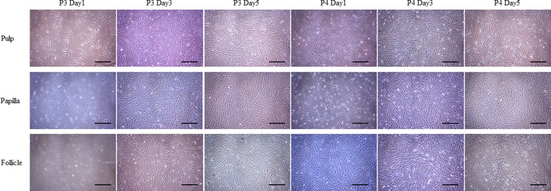
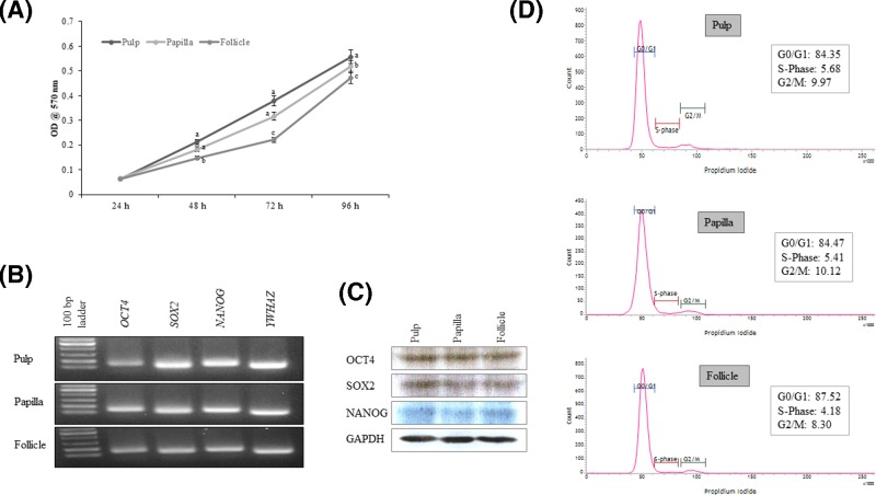
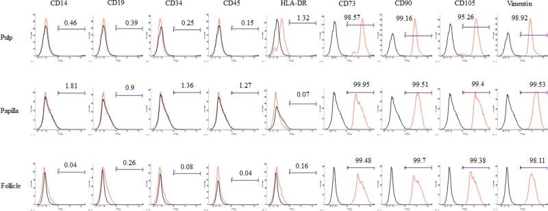
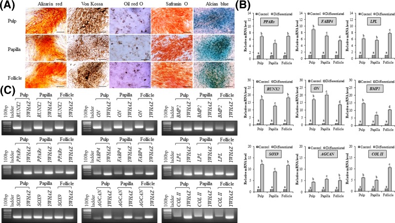
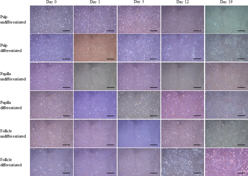
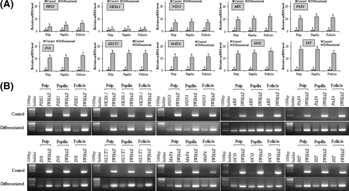
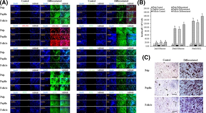
The present study was carried out to investigate and compare the in vitro differentiation potential of mesenchymal stem cells (MSCs) isolated from human dental tissues (pulp, papilla, and follicle) of the same donor. MSCs were isolated from dental tissues (pulp, papilla, and follicle) following digestion method and were analyzed for the expression of pluripotent markers and cell surface markers. All three types of MSCs were evaluated for their potential to differentiate into mesenchymal lineages. Further, the MSCs were differentiated into pancreatic β cell-like cells using multistep protocol and characterized for the expression of pancreatic lineage specific markers. Functional properties of differentiated pancreatic β cell-like cells were assessed by dithizone staining and glucose challenge test. All three types of MSCs showed fibroblast-like morphology upon culture and expressed pluripotent, and mesenchymal cell surface markers. These MSCs were successfully differentiated into mesenchymal lineages and transdifferentiated into pancreatic β cell-like cells. Among them, dental follicle derived MSCs exhibits higher transdifferentiation potency toward pancreatic lineage as evaluated by the expression of pancreatic lineage specific markers both at mRNA and protein level, and secreted higher insulin upon glucose challenge. Additionally, follicle-derived MSCs showed higher dithizone staining upon differentiation. All three types of MSCs from a single donor possess similar cellular properties and can differentiate into pancreatic lineage. However, dental follicle derived MSCs showed higher potency toward pancreatic lineage than pulp and papilla derived MSCs, suggesting their potential application in future stem cell based therapy for the treatment of diabetes.

Keywords: Diabetes; Dithizone; Glucose challenge; MSCs; Pancreatic lineage; Pluripotency.


Conflict of interest statement

The authors declare that there are no competing interests associated with the manuscript.

Figures

Figure 1
Figure 1
Phase contrast microscopic images MSCs isolated from dental tissues such as pulp, papilla, and follicle showed adherent fibroblast-like morphology at different time intervals at passages P3 and P4; scale bar = 100 μm.
Figure 2
Figure 2
Characterization of MSCs isolated from dental tissues such as pulp, papilla, and follicle (A) Analysis of cell proliferation using MTT assay. Data are represented for each sample conducted in triplicates from three independent experiments. Significant differences are indicated using different letters when P<0.05. (B) Analysis of the expression of pluripotent marker genes using polymerase chain reaction that was evaluated by agarose gel electrophoresis. All three types of MSCs expressed octamer-binding transcription factor 4 (OCT4), sex determining region Y-box 2 (SOX2), and NANOG. (C) Western blot images showing the expression of pluripotent marker proteins such as OCT4, SOX2, and NANOG in MSCs isolated from pulp, papilla, and follicle dental tissues. (D) Flow cytometric analysis of three types of MSCs at passage 3 demonstrating normal DNA content in gap0/1 (G0/G1), synthesis (S), or gap2/mitotic (G2/M) phases of the cell cycle. A total of 10,000 MSCs were counted for each sample in triplicates from three independent experiments.
Figure 3
Figure 3
Flow cytometric analysis of the expression of cell surface markers in MSCs isolated from pulp, papilla, and follicle dental tissues All three types of MSCs were positive for the expression of mesenchymal markers such as CD73, CD90, CD105, and internal marker vimentin, whereas these cells were negative for the expression of monocyte marker CD14, B-lymphocyte marker CD19, HLA-DR, and hematopoietic markers CD34 and CD45.
Figure 4
Figure 4
Analysis of in vitro mesenchymal lineage differentiation (A) MSCs isolated from dental pulp, papilla, and follicle tissues were differentiated into mesenchymal lineages such as osteocytes, adipocytes, and chondrocytes. The differentiation was confirmed by lineage-specific staining (osteocytes: alizarin red and von Kossa; adipocytes: oil red O; chondrocytes: safranin O and alcian blue); scale bar = 100 μm. (B) RT-qPCR analysis of mRNA expression of mesenchymal lineage specific marker genes. The relative mRNA level was quantified using 2−∆∆CT method using tyrosine 3-monooxygenase/tryptophan 5-monooxygenase activation protein, zeta polypeptide (YWHAZ) for normalization. The mRNA levels are expressed as fold change in relation to the undifferentiated control MSCs [peroxisome proliferator-activated receptor γ-2 (PPARγ), fatty-acid binding protein 4 (FABP4), and lipoprotein lipase (LPL), adipocyte-specific; runt-related transcription factor 2 (RUNX2), osteonectin (ON), and bone morphogenetic protein 2 (BMP2), osteocyte-specific; and sex determining region Y-box 9 (SOX9), cartilage-specific proteoglycan core protein (AGGRECAN), and type II collagen (COLLAGEN II), chondrocyte-specific]. Data represent the mean ± SE obtained in triplicates from three independent experiments. Significant differences are shown using different letters when P<0.05. (C) The product size of each marker genes was evaluated by agarose gel electrophoresis.
Figure 5
Figure 5
Phase contrast images showing morphological changes occurred during in vitro differentiation of MSCs isolated from dental pulp, papilla, and follicle tissues into pancreatic β cell-like cells All three types of MSCs were successfully differentiated toward pancreatic lineage and showed change in morphologies at different stages of differentiation; scale bar = 100 μm.
Figure 6
Figure 6
Analysis of the expression of pancreatic lineage specific marker genes after differentiation (A) RT-qPCR analysis for the evaluation of fold change in the expression of pancreatic lineage specific marker genes such as pancreatic duodenal homeobox-1 (PDX-1), NK6 homeobox 1 (NKX6.1), neurogenin 3 (NGN3), aristaless-related homeobox (ARX), paired box 4 (PAX4), insulin (INS), solute carrier family 2 member 2 (GLUT2), MAF bZIP transcription factor A (MAFA), glucagon (GCG), and somatostatin (SST). The expression levels are expressed as change in relative mRNA level in relation to the undifferentiated control using 2−∆∆CT method. Tyrosine 3-monooxygenase/tryptophan 5-monoxygenase activation protein, zeta polypeptide (YWHAZ) was used for data normalization. The data are represented as mean ± SE obtained in triplicates from three independent experiments. Significant differences are denoted using different letters when P<0.05. (B) The product size of each marker genes was evaluated by agarose gel electrophoresis.
Figure 7
Figure 7
Immunocytochemistry and functional evaluation of differentiated pancreatic β cell-like cells (A) Analysis of the expression of pancreatic lineage specific marker proteins by immunocytochemistry; scale bar = 100 μm. (B) Evaluation of insulin secretion by glucose challenge test. The data are represented as mean ± SE obtained in triplicates from three independent experiments. Significant differences are denoted using different letters when P<0.05. (C) Differentiated cells were evaluated for the presence of zinc ions by staining with dithizone; scale bar = 100 μm.

Similar articles

Cited by

References

    1. Wild S., Roglic G., Green A.. et al. (2004) Global prevalence of diabetes: estimates for the year 2000 and projections for 2030. Diabetes Care 27, 1047–1053 10.2337/diacare.27.5.1047 - DOI - PubMed
    1. Bouwens L., Houbracken I. and Mfopou J.K. (2013) The use of stem cells for pancreatic regeneration in diabetes mellitus. Nat. Rev. Endocrinol. 9, 598–606 10.1038/nrendo.2013.145 - DOI - PubMed
    1. Godfrey K.J., Mathew B., Bulman J.C.. et al. (2012) Stem cell-based treatments for Type 1 diabetes mellitus: bone marrow, embryonic, hepatic, pancreatic and induced pluripotent stem cells. Diabetes Med. 29, 14–23 10.1111/j.1464-5491.2011.03433.x - DOI - PubMed
    1. Lilly M.A., Davis M.F., Fabie J.E.. et al. (2016) Current stem cell based therapies in diabetes. Am. J. Stem Cells. 5, 87–98 - PMC - PubMed
    1. Xin Y., Jiang X., Wang Y.. et al. (2016) Insulin-producing cells differentiated from human bone marrow mesenchymal stem cells in vitro ameliorate streptozotocin-induced diabetic hyperglycemia. PLoS One 11, e0145838 10.1371/journal.pone.0145838 - DOI - PMC - PubMed

Publication types

Substances