

LncRNA TP73-AS1 down-regulates miR-139-3p to promote retinoblastoma cell proliferation
- PMID: 31015368
- PMCID: PMC6509052
- DOI: 10.1042/BSR20190475
LncRNA TP73-AS1 down-regulates miR-139-3p to promote retinoblastoma cell proliferation
Abstract
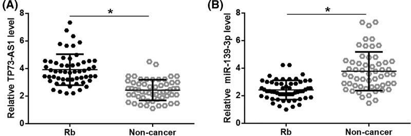
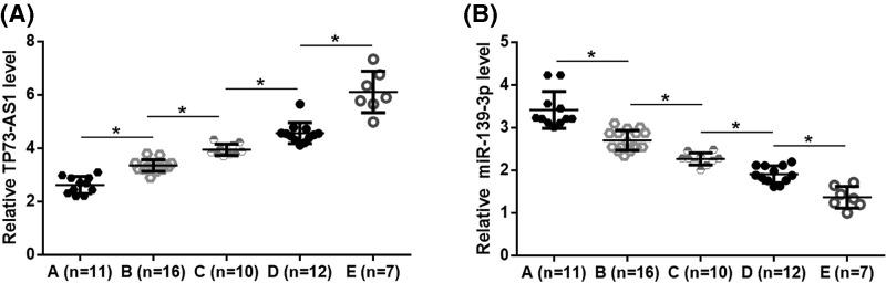
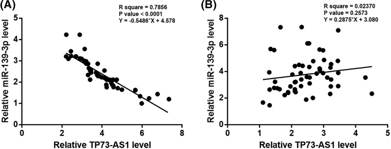
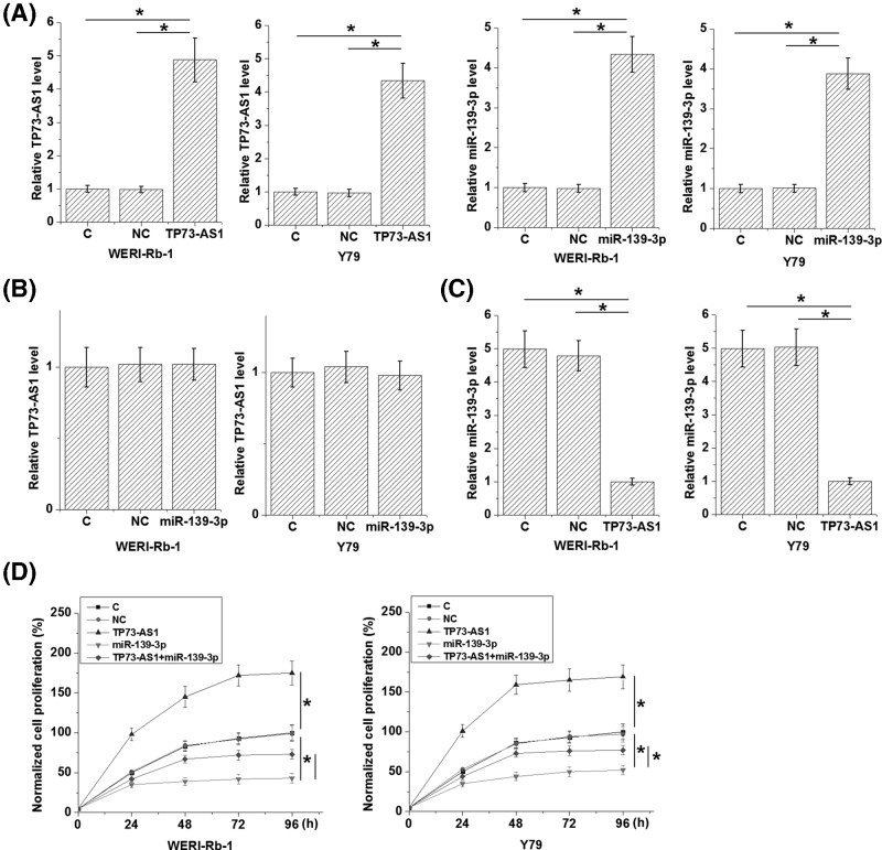
Our study aimed to investigate the role of long non-coding RNAs (lncRNA) TP73-AS1 in retinoblastoma (Rb). In the present study, we found that TP73-AS1 was up-regulated, while miR-139-3p was down-regulated in Rb. TP73-AS1 and miR-139-3p were inversely correlated in Rb tissues. In cells of Rb cell lines, overexpression of miR-139-3p failed to affect TP73-AS1, while TP73-AS1 overexpression caused the down-regulated miR-139-3p TP73-AS1 overexpression caused promoted proliferation of Rb cells but showed no significant effects on cell migration and invasion. miR-139-3p overexpression played an opposite role and attenuated the effects of TP73-AS1 overexpression. Therefore, lncRNA TP73-AS1 may down-regulate miR-139-3p to promote Rb cell proliferation.
Keywords: lncRNA TP73-AS1; miR-139-3p; proliferation; retinoblastoma.
© 2019 The Author(s).
Conflict of interest statement
The authors declare that there are no competing interests associated with the manuscript.
Figures
Similar articles
-
The long non-coding RNA ILF3-AS1 increases the proliferation and invasion of retinoblastoma through the miR-132-3p/SMAD2 axis.Exp Cell Res. 2020 Aug 15;393(2):112087. doi: 10.1016/j.yexcr.2020.112087. Epub 2020 May 11. Exp Cell Res. 2020. PMID: 32407730
-
LncRNA TMPO-AS1 up-regulates the expression of HIF-1α and promotes the malignant phenotypes of retinoblastoma cells via sponging miR-199a-5p.Pathol Res Pract. 2020 Apr;216(4):152853. doi: 10.1016/j.prp.2020.152853. Epub 2020 Feb 5. Pathol Res Pract. 2020. PMID: 32139259
-
Long Noncoding RNA TP73-AS1 Targets MicroRNA-329-3p to Regulate Expression of the SMAD2 Gene in Human Cervical Cancer Tissue and Cell Lines.Med Sci Monit. 2019 Oct 30;25:8131-8141. doi: 10.12659/MSM.916292. Med Sci Monit. 2019. Retraction in: Med Sci Monit. 2023 Dec 25;29:e943586. doi: 10.12659/MSM.943586. PMID: 31663517 Free PMC article. Retracted.
-
A review on the role of oncogenic lncRNA OIP5-AS1 in human malignancies.Biomed Pharmacother. 2021 May;137:111366. doi: 10.1016/j.biopha.2021.111366. Epub 2021 Feb 15. Biomed Pharmacother. 2021. PMID: 33601149 Review.
-
Long non-coding RNA TP73-AS1 in cancers.Clin Chim Acta. 2020 Apr;503:151-156. doi: 10.1016/j.cca.2019.12.025. Epub 2020 Jan 21. Clin Chim Acta. 2020. PMID: 31978409 Review.
Cited by
-
Bioinformatics analysis proposes a possible role for long noncoding RNA MIR17HG in retinoblastoma.Cancer Rep (Hoboken). 2024 Feb;7(2):e1933. doi: 10.1002/cnr2.1933. Epub 2024 Feb 6. Cancer Rep (Hoboken). 2024. PMID: 38321787 Free PMC article.
-
circKIF4A promotes tumorogenesis of glioma by targeting miR-139-3p to activate Wnt5a signaling.Mol Med. 2020 Apr 8;26(1):29. doi: 10.1186/s10020-020-00159-1. Mol Med. 2020. PMID: 32268875 Free PMC article.
-
Long non-coding RNA BCAR4 aggravated proliferation and migration in esophageal squamous cell carcinoma by negatively regulating p53/p21 signaling pathway.Bioengineered. 2021 Dec;12(1):682-696. doi: 10.1080/21655979.2021.1887645. Bioengineered. 2021. PMID: 33602031 Free PMC article.
-
A predictive analysis approach for paediatric and adult high-grade glioma: miRNAs and network insight.Ann Transl Med. 2020 Mar;8(5):242. doi: 10.21037/atm.2020.01.12. Ann Transl Med. 2020. PMID: 32309389 Free PMC article.
-
Role of non-coding RNAs and exosomal non-coding RNAs in retinoblastoma progression.Front Cell Dev Biol. 2022 Dec 23;10:1065837. doi: 10.3389/fcell.2022.1065837. eCollection 2022. Front Cell Dev Biol. 2022. PMID: 36619866 Free PMC article. Review.
References
Publication types
MeSH terms
Substances
LinkOut - more resources
Full Text Sources