Review
. 2019 Apr 23;63(1):29-43.
doi: 10.1042/EBC20180055. Print 2019 Apr 23.

The histone chaperoning pathway: from ribosome to nucleosome

Affiliations
Review

The histone chaperoning pathway: from ribosome to nucleosome

Alonso J Pardal et al. Essays Biochem. .

Abstract

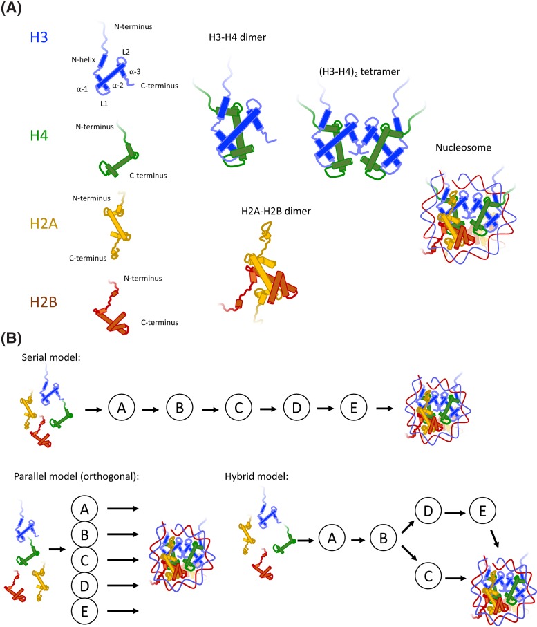
Nucleosomes represent the fundamental repeating unit of eukaryotic DNA, and comprise eight core histones around which DNA is wrapped in nearly two superhelical turns. Histones do not have the intrinsic ability to form nucleosomes; rather, they require an extensive repertoire of interacting proteins collectively known as 'histone chaperones'. At a fundamental level, it is believed that histone chaperones guide the assembly of nucleosomes through preventing non-productive charge-based aggregates between the basic histones and acidic cellular components. At a broader level, histone chaperones influence almost all aspects of chromatin biology, regulating histone supply and demand, governing histone variant deposition, maintaining functional chromatin domains and being co-factors for histone post-translational modifications, to name a few. In this essay we review recent structural insights into histone-chaperone interactions, explore evidence for the existence of a histone chaperoning 'pathway' and reconcile how such histone-chaperone interactions may function thermodynamically to assemble nucleosomes and maintain chromatin homeostasis.

Keywords: Chaperone; HIstone chaperone; Nucleosome; chromatin; histones.


Conflict of interest statement

The authors declare that there are no competing interests associated with the manuscript.

Figures

Figure 1
Figure 1. Components of the nucleosome and considerations for multichaperone networks
(A) Nucleosomes comprise of four core histones – H2A, H2B, H3 and H4. The central histone fold comprises three helices, α1, α2 and α3, which can be flanked by extension on the N and C termini. N-terminal extensions are in the form of basic tails, and in the case of H3, an additional α-helix (αN-helix). H3 and H4 fold to form an H3–H4 dimer, two of which can form an (H3–H4)2-heterotetramer. The tetramer adopts the central location in the nucleosome, known as a ‘tetrasome’ when bound to DNA on its own, and is capped by two H2A–H2B dimers to form the nucleosome core particle, which wraps 145–147 base pairs of DNA in almost two superhelical turns. (B) Histones associate with a large repertoire of chaperoning complexes, represented as A, B, C, D and E. Assuming mutual exclusivity in binding, histones may transition through multiple chaperoning complexes before their incorporation in chromatin (a serial pathway), or each chaperoning complex may represent an orthologous route to deposition (a parallel pathway), or indeed, a mixture of the two (a hybrid model). In reality, a hybrid model which bifurcates depending of histone isotype best explains the H3–H4 deposition pathway (see Figure 2).
Figure 2
Figure 2. Histone synthesis and nuclear import
After synthesis histones H3 and H4 interact in the cytosol with common folding chaperones HSC70 and HSP90, with H3 being mono-methylated on K9 co-translationally by SetDB1. Two pathways have been proposed for the import of H3 and H4: folding of an H3–H4 dimer in the cytosol and subsequent binding to ASF1 before import by IPO4, or import of monomeric histones bound directly to importins, with H3–H4 dimerisation occurring in the nucleus. ASF1’s interaction with H3–H4 in the cytosol can be regulated by the protein CDAN1, which competes with histone for ASF1 binding. H2A and H2B are imported as dimers bound to NAP1 and IPO9. In the nucleus, NASP interacts with H3 downstream of ASF1 through a high affinity interaction with the H3 α3 helix, and may thereby function as a receptor of H3 from the import machinery. Whether a monomeric H4 chaperone exists is not known. RbAp46 and HAT1 associate with H4, acetylating K5 and K12, and together with NASP and ASF1 form the major soluble H3–H4 complex. The S-phase-specific TLK1 and TLK2 can increase the affinity between histones and ASF1 through ASF1-specific phosphorylation marks.
Figure 3
Figure 3. Histone chaperoning in the nucleus
Histone deposition can be split into replication dependent, and replication independent mechanisms. The variants H3.1/H3.2–H4 are incorporated into chromatin predominantly at the replication fork during replication dependent deposition, in which a number of chaperoning components have been implicated, including FACT (SpT16-SSRP1), CAF1 (p150, p60, RbAp48), and the replication proteins POLE3–4, RPA, MCM2 (N-terminal tail) and TONSL. TONSL is also involved in the DNA damage response, a role that may be related to its chromatin assembly function. ASF1 may act as a bridging factor between soluble pool and deposition factors. In addition to H3–H4, the FACT complex also binds to H2A–H2B through its U-turn motif and C-terminal acidic stretch, and may be responsible for governing H2A–H2B deposition post replication, or indeed, the chaperones NAP1 and Nucleoplasmin may play a role. Replication independent deposition occurs throughout the cell cycle, utilising the H3.3 variant. At sites of transcription both the HIRA complex (HIRA, UBN1 and CABIN1), which can bind naked DNA, and FACT complex, which associates with elongating RNA pol II, are involved in H3–H4 deposition, with the FACT complex also extending its role to H2A–H2B incorporation. At other sites in the genome, including heterochromatic repetitive elements, the chaperone DAXX and chromatin remodeller ATRX govern the incorporation of H3.3. Histone chaperones also play a central role during DNA repair, omitted here for brevity. For a detailed description we refer the reader to some recent excellent reviews [131–133].
Figure 4
Figure 4. Transitioning through the thermodynamic landscape of nucleosome assembly
Histones must transition through a number of protein complexes in order to fold with DNA into nucleosomes without the input of energy from ATP hydrolysis. Immediately after synthesis histones contain a high free energy. This energy may be captured by histone chaperones and utilised in a way which drives correct folding and oligomerisation of histone subunits. This is mostly likely achieved through extensive and specific interactions covering all transitions states of histone intermediates. In such a scenario, accepting potential ATP-driven input from canonical protein folding chaperones in the initial stages, the histone chaperoning pathway may represent an efficient way to assemble a highly abundant cellular complex.

Similar articles

Cited by

References

    1. Hübner M.R., Eckersley-Maslin M.A. and Spector D.L. (2013) Chromatin organization and transcriptional regulation. Curr. Opin. Genet. Dev. 23, 89–95 10.1016/j.gde.2012.11.006 - DOI - PMC - PubMed
    1. Mondal T., Rasmussen M., Pandey G.K., Isaksson A. and Kanduri C. (2010) Characterization of the RNA content of chromatin. Genome Res. 20, 899–907 10.1101/gr.103473.109 - DOI - PMC - PubMed
    1. Luger K., Mäder A.W., Richmond R.K., Sargent D.F. and Richmond T.J. (1997) Crystal structure of the nucleosome core particle at 2.8 A resolution. Nature 389, 251–260 10.1038/38444 - DOI - PubMed
    1. Arents G. and Moudrianakis E.N. (1995) The histone fold: a ubiquitous architectural motif utilized in DNA compaction and protein dimerization. Proc. Natl. Acad. Sci. U. S. A. 92, 11170–11174 10.1073/pnas.92.24.11170 - DOI - PMC - PubMed
    1. Davey C.A., Sargent D.F., Luger K., Maeder A.W. and Richmond T.J. (2002) Solvent mediated interactions in the structure of the nucleosome core particle at 1.9 A resolution. J. Mol. Biol. 319, 1097–1113 10.1016/S0022-2836(02)00386-8 - DOI - PubMed

Publication types