. 2019 Apr 23;10(1):1849.
doi: 10.1038/s41467-019-09507-0.

Structural insight into YcbB-mediated beta-lactam resistance in Escherichia coli

Affiliations

Structural insight into YcbB-mediated beta-lactam resistance in Escherichia coli

Nathanael A Caveney et al. Nat Commun. .

Abstract

The bacterial cell wall plays a crucial role in viability and is an important drug target. In Escherichia coli, the peptidoglycan crosslinking reaction to form the cell wall is primarily carried out by penicillin-binding proteins that catalyse D,D-transpeptidase activity. However, an alternate crosslinking mechanism involving the L,D-transpeptidase YcbB can lead to bypass of D,D-transpeptidation and beta-lactam resistance. Here, we show that the crystallographic structure of YcbB consists of a conserved L,D-transpeptidase catalytic domain decorated with a subdomain on the dynamic substrate capping loop, peptidoglycan-binding and large scaffolding domains. Meropenem acylation of YcbB gives insight into the mode of inhibition by carbapenems, the singular antibiotic class with significant activity against L,D-transpeptidases. We also report the structure of PBP5-meropenem to compare interactions mediating inhibition. Additionally, we probe the interaction network of this pathway and assay beta-lactam resistance in vivo. Our results provide structural insights into the mechanism of action and the inhibition of L,D-transpeptidation, and into YcbB-mediated antibiotic resistance.


Conflict of interest statement

The authors declare no competing interests.

Figures
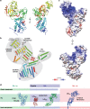
Fig. 1
Fig. 1
Overall architecture of E. coli YcbB. a The crystal structure of YcbB-meropenem acyl-enzyme complex in ribbon representation, coloured in rainbow from N-terminus (blue) to C-terminus (red). Meropenem stick representation is coloured in black and by heteroatom. Two views related by a 90° rotation along the y-axis. b Topology diagram of YcbB-meropenem acyl-enzyme complex, coloured as in a. The catalytic, PG and scaffolding domains are annotated and circled in grey. c Electrostatic surface representation of YcbB in two views related by a 180° rotation around the y-axis. d Diversity of structurally characterised L,D-transpeptidases. Gram negative (green background), soluble YcbB in sky blue; Mycobacterial (blue background), lipoprotein LdtMt1 (PDBID 5E5L) and LdtMt2 (PDBID 5DU7) in red and green; Gram positive (red background), TM anchored Ldtfm (1ZAT) in dark blue
Fig. 2
Fig. 2
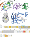
Structure of the E. coli YcbB peptidoglycan binding domain. a Ribbon representation of the E. coli YcbB peptidoglycan binding domain, residues 241–351, with residue conservation mapped to backbone colour from red to blue in decreasing conservation. Conserved core residues labelled in black and key, surface exposed, peptidoglycan binding residues labelled in red. b Overlay of YcbB peptidoglycan binding domain and high scoring peptidoglycan binding domains from a structural homology search on the Dali server. YcbB in red, gp144, a Pseudomonas aeruginosa phage phiKZ endolysin in salmon (PDB ID 3BKH), a Clostridium acetobutylicum zinc-dependent amidase in white (PDB ID 4XXT), a Clostridium difficile PBP in pale blue (PDB ID 5TV7), and gp15, a Burkholderia AP3 phage endolysin in dark blue (PDB ID 5NM7). Peptidoglycan binding domain structures are coloured from red to blue based on decreasing structural homology. c The sequence of E. coli YcbB (YcbB_Ec) aligned with peptidoglycan binding domains from phiKZ gp144 (gp144_phiKZ), C. acetobutylicum zinc-dependent amidase (Zn_amidase_Ca), C. difficile PBP (PBP_Cd), and AP3 gp15 (gp15_AP3). Secondary structure of E. coli YcbB is displayed atop the sequence alignment, with the extended loop shown in red and the unstructured region in grey
Fig. 3
Fig. 3
Structure of the E. coli YcbB catalytic domain. a A ribbon representation of the YcbB catalytic domain, residues 375–576, coloured in rainbow from red (N-terminus) to blue (C-terminus). Meropenem is represented in black and coloured by heteroatom. The unique capping sub-domain is highlighted in a bold silhouette. b The catalytic domain architecture of Enterococcus faecium LdtFm (PDB ID 3ZGP) and Bacillus subtilis YkuD (PDB ID 1Y7M) highlighting the differences seen among L,D-transpeptidase catalytic domains in the capping loop region. c Cut surface representation of the YcbB catalytic domain, showing the capped cleft formed between the donor and acceptor sites by the capping loop sub-domain. The catalytic Cys528 and His509 are coloured in yellow and blue respectively. Meropenem (blue) is overlaid in its position in the donor site, acylating the catalytic cysteine. d The active site architecture of E. coli YcbB. The catalytic Cys528 is acylated by meropenem and the sulphide is rotated away from the Nτ of His509 (4.2 Å) which activated the cystine prior to acylation. The Nπ of His509 is seen to be in close proximity with the backbone carbonyl of the adjacent Asp510 (2.7 Å), stabilising and promoting formation of the histidine cation. e The sequence of E. coli YcbB (Ec_YcbB) aligned with the aforementioned catalytic domains from E. faecium and B. subtilis (Ef_Ldtfm and Bs_Yku, respectively) as well as LdtMt1 and LdtMt2 from Mycobacterium tuberculosis (Mt_LdtMt1 and Mt_LdtMt2, respectively)
Fig. 4
Fig. 4
Meropenem inhibition of E. coli YcbB and PBP5. a Chemical diagram of meropenem pre- and post-acylation by YcbB and PBP5. b mFo-DFc simulated annealing omit map for the acylated meropenem on Cys528 of YcbB, contoured at 2σ. YcbB is in blue and meropenem in grey. The catalytic dyad (Cys528 and His509), selected residues, and meropenem are coloured by heteroatom. Hydrogen bonding between meropenem and the backbone carbonyl of Ala505 is represented with dashed lines. c mFo-DFc simulated annealing omit map for the acylated meropenem on Ser73 of PBP5, contoured at 2σ. PBP5 is in blue and meropenem in grey. PBP5 residues involved in the stabilization of meropenem, relevant water molecules, and meropenem are coloured by heteroatom. Hydrogen bonding between meropenem, selected residues, and water is represented with dashed lines. Relative arrangement of nucleophile, general base and oxyanion hole (backbone atoms shown) for PBP5 (d) and YcbB (e). f, g LigPlot diagrams of meropenem acyl-enzyme complexes. f YcbB-meropenem complex with meropenem in grey and protein residues in white, with atoms coloured by heteroatom. g PBP5-meropenem complex coloured as in f. h Overlay of meropenem-acylated catalytic residues of YcbB (darker blue and green) and PBP5 (lighter blue and pink) showing a similar general curvature of both meropenem molecules, with the notable exception of an approximate 180° rotation about meropenem C6 due to the differing active site architectures of YcbB and PBP5
Fig. 5
Fig. 5
In vivo assay of YcbB mediated beta-lactam resistance. a Various replacements and deletions of the catalytic residues (in red), capping loop region (in green) and PG binding domain (in blue), mapped onto the catalytic and PG domains of YcbB. Replacements retaining YcbB mediated beta-lactam (ampicillin and ceftriaxone) resistance are denoted as β-lacR, while replacements and deletions resulting in beta-lactam (ampicillin and ceftriaxone) susceptibility are denoted as β-lacS. b Replacements and deletions of pTK2(ycbB) (i–viii) are listed with their resulting phenotype
Fig. 6
Fig. 6
Schematic representation of YcbB mediated beta-lactam resistance. ae Assembly and function of the YcbB mediated beta-lactam resistance pathway. a PBP1b function in the absence of beta-lactam antibiotics. PBP1b acts as both glycosyltransferase and D,D-transpeptidase. b PBP1b function in the presence of beta-lactam antibiotics, such as ampicillin. PBP1b can maintain glycosyltransferase activity, but D,D-transpeptidation is inhibited. c Beta-lactam resistance complex formation upon production of alarmone. YcbB likely interacts with PBP1b and PBP5 through its putative interaction/helical domain. d YcbB is likely additionally associated and oriented through its PG binding domain interacting with the acceptor strand of the existing PG sacculus. e The resistance complex can successfully rescue crosslinking function. PBP1b polymerizes PG strands with glycosyltransferase activity, PBP5 removed a terminal D-alanine with its carboxypeptidase activity, and YcbB acts on the modified peptides with L,D-transpeptidase activity to crosslink the PG strand into the sacculus. fj Function and mechanism of the YcbB catalytic domain during L,D-transpeptidation. f Tetrapeptide of the donor PG strand enters the donor site of the catalytic domain. g Donor tetrapeptide is acylated at its meso-DAP residue by the catalytic cysteine, resulting in the loss of the terminal D-alanine. h Adjacent acceptor PG peptide enters the acceptor site of the catalytic domain. i The covalent acyl-enzyme is deacylated via nucleophilic attack of the side chain meso-DAP on the adjacent acceptor PG strand, forming the L,D-crosslink. j Rearrangement of the capping loop/sub-domain from a capped conformation to an open cleft will occur, resulting in the release of the crosslinked substrate

Similar articles

Cited by

References

    1. Sobhanifar S, King DT, Strynadka NCJ. Fortifying the wall: synthesis, regulation and degradation of bacterial peptidoglycan. Curr. Opin. Struct. Biol. 2013;23:695–703. doi: 10.1016/j.sbi.2013.07.008. - DOI - PubMed
    1. Lovering AL, Safadi SS, Strynadka NCJ. Structural Perspective of Peptidoglycan Biosynthesis and Assembly. Annu. Rev. Biochem. 2012;81:451–478. doi: 10.1146/annurev-biochem-061809-112742. - DOI - PubMed
    1. Caveney NA, Li FK, Strynadka NC. Enzyme structures of the bacterial peptidoglycan and wall teichoic acid biogenesis pathways. Curr. Opin. Struct. Biol. 2018;53:45–58. doi: 10.1016/j.sbi.2018.05.002. - DOI - PubMed
    1. King DT, Sobhanifar S, Strynadka NCJ. One ring to rule them all: current trends in combating bacterial resistance to the β-lactams. Protein Sci. 2016;25:787–803. doi: 10.1002/pro.2889. - DOI - PMC - PubMed
    1. Hugonnet JE, et al. Factors essential for L,D-transpeptidase-mediated peptidoglycan cross-linking and β-lactam resistance inEscherichia coli. eLife. 2016;5:1–22. doi: 10.7554/eLife.19469. - DOI - PMC - PubMed

Publication types

MeSH terms

LinkOut - more resources