. 2019 Apr 23;9(1):6438.
doi: 10.1038/s41598-019-42896-2.

Phenotypic characteristics and transcriptome profile of Cryptococcus gattii biofilm

Affiliations

Phenotypic characteristics and transcriptome profile of Cryptococcus gattii biofilm

Eliandro Reis Tavares et al. Sci Rep. .

Abstract

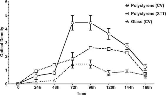
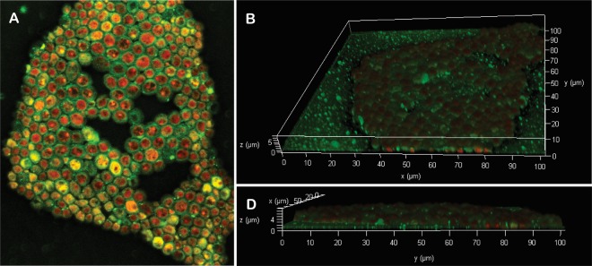
In this study, we characterized Cryptococcus gattii biofilm formation in vitro. There was an increase in the density of metabolically active sessile cells up to 72 h of biofilm formation on polystyrene and glass surfaces. Scanning electron microscopy and confocal laser scanning microscopy analysis revealed that in the early stage of biofilm formation, yeast cells adhered to the abiotic surface as a monolayer. After 12 h, extracellular fibrils were observed projecting from C. gattii cells, connecting the yeast cells to each other and to the abiotic surface; mature biofilm consisted of a dense network of cells deeply encased in an extracellular polymeric matrix. These features were also observed in biofilms formed on polyvinyl chloride and silicone catheter surfaces. We used RNA-Seq-based transcriptome analysis to identify changes in gene expression associated with C. gattii biofilm at 48 h compared to the free-floating planktonic cells. Differential expression analysis showed that 97 and 224 transcripts were up-regulated and down-regulated in biofilm, respectively. Among the biological processes, the highest enriched term showed that the transcripts were associated with cellular metabolic processes, macromolecule biosynthetic processes and translation.


Conflict of interest statement

The authors declare no competing interests.

Figures

Figure 1
Figure 1
Biofilm formation by clinical (n = 4) and environmental (n = 4) isolates of Cryptococcus gattii complex, and Cryptococcus gatti ATCC 24065 reference strain on polystyrene surface incubated in Sabouraud dextrose broth for 72 h at 37 °C. Biofilm biomass was measured after crystal violet staining (OD570nm); the values represent the mean ± standard deviation and are representative of three independent experiments. Clinical isolates: 840244, LCF-312, 62752 and CG03; Environmental isolates: 3A1, 2B4, 1A2 and 3A4. Bars not sharing a letter differ significantly (P < 0.05) between the isolates.
Figure 2
Figure 2
Temporal development of Cryptococcus gattii ATCC 24065 biofilm on polystyrene and glass surfaces monitored by measuring the biomass of sessile cells using crystal violet (CV) staining (OD570nm) and XTT reduction (OD492nm) methods. The values represent the mean ± SD and are representative of three independent experiments.
Figure 3
Figure 3
Scanning electron microscopy (SEM) images of Cryptococcus gattii ATCC 24065 biofilm formation stages on glass surface over a period of time of incubation at 37 °C. A gradual increase in both, the cell number and the amount of fibrils was observed over time. (A) 12 hours; (B) 24 hours; (C) 48 hours; (D) 72 hours; (E) 96 hours; and (F) 120 hours.
Figure 4
Figure 4
Confocal laser scanning microscopy (CLSM) images of the Cryptococcus gattii ATCC 24065 biofilm formed on glass surface after 72 h at 37 °C. Mature biofilm consisted of metabolically active (red-fluorescence due to FUN-1 staining) sessile cells encased in an extracellular polysaccharide-like substance (green-fluorescence due to Con-A staining) organized in an 8-µm-thick monolayer. (A) Panoramic view of biofilm; (B,C) Three-dimensional biofilm reconstitution.
Figure 5
Figure 5
Validation of the data generated by RNA-seq with relative quantitative real-time PCR analysis. mRNAs from planktonic and 48 h-biofilm cells of Cryptococcus gattii complex were obtained and the expression of four selected differentially expressed genes in biofilm was quantified by real-time PCR using the QuantiNova SYBR Green RT-PCR system and the cycle threshold method. Changes in transcript levels were determined using ACT gene, coding for actin, (Access number: XM_003191370) as an internal control. Results are the mean ± standard error for duplicate determinations and are representative of three independent experiments. Results are presented as the relative gene expression of selected genes related to the control (line). Significant differences were observed between the planktonic and biofilm cells (P < 0.05).

Similar articles

Cited by

References

    1. Donlan Rodney M. Biofilms: Microbial Life on Surfaces. Emerging Infectious Diseases. 2002;8(9):881–890. doi: 10.3201/eid0809.020063. - DOI - PMC - PubMed
    1. Donlan RM, Costerton JW. Biofilms: survival mechanisms of clinically relevant microorganisms. Clin. Microbiol. Rev. 2002;15:167–93. doi: 10.1128/CMR.15.2.167-193.2002. - DOI - PMC - PubMed
    1. Martinez LR, Casadevall A. Cryptococcus neoformans Biofilm Formation Depends on Surface Support and Carbon Source and Reduces Fungal Cell Susceptibility to Heat, Cold, and UV Light. Appl. Environ. Microbiol. 2007;73:4592–4601. doi: 10.1128/AEM.02506-06. - DOI - PMC - PubMed
    1. Ravi S, Pierce C, Witt C, Wormley FL. Biofilm formation by Cryptococcus neoformans under distinct environmental conditions. Mycopathologia. 2009;167:307–314. doi: 10.1007/s11046-008-9180-6. - DOI - PMC - PubMed
    1. Benaducci, T. et al. Virulence of Cryptococcus sp. Biofilms In Vitro and In Vivo using Galleria mellonella as an Alternative Model. Front. Microbiol. 7 (2016). - PMC - PubMed

Publication types

MeSH terms