. 2018 Dec;2(12):930-941.
doi: 10.1038/s41551-018-0271-5. Epub 2018 Jul 23.

A tissue-engineered scale model of the heart ventricle

Affiliations

A tissue-engineered scale model of the heart ventricle

Luke A MacQueen et al. Nat Biomed Eng. 2018 Dec.

Erratum in

  • Addendum: A tissue-engineered scale model of the heart ventricle.
    MacQueen LA, Sheehy SP, Chantre CO, Zimmerman JF, Pasqualini FS, Liu X, Goss JA, Campbell PH, Gonzalez GM, Park SJ, Capulli AK, Ferrier JP, Kosar TF, Mahadevan L, Pu WT, Parker KK. MacQueen LA, et al. Nat Biomed Eng. 2022 Nov;6(11):1318. doi: 10.1038/s41551-022-00854-w. Nat Biomed Eng. 2022. PMID: 35260798 No abstract available.

Abstract

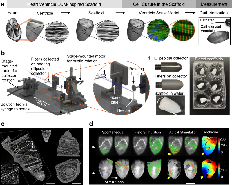
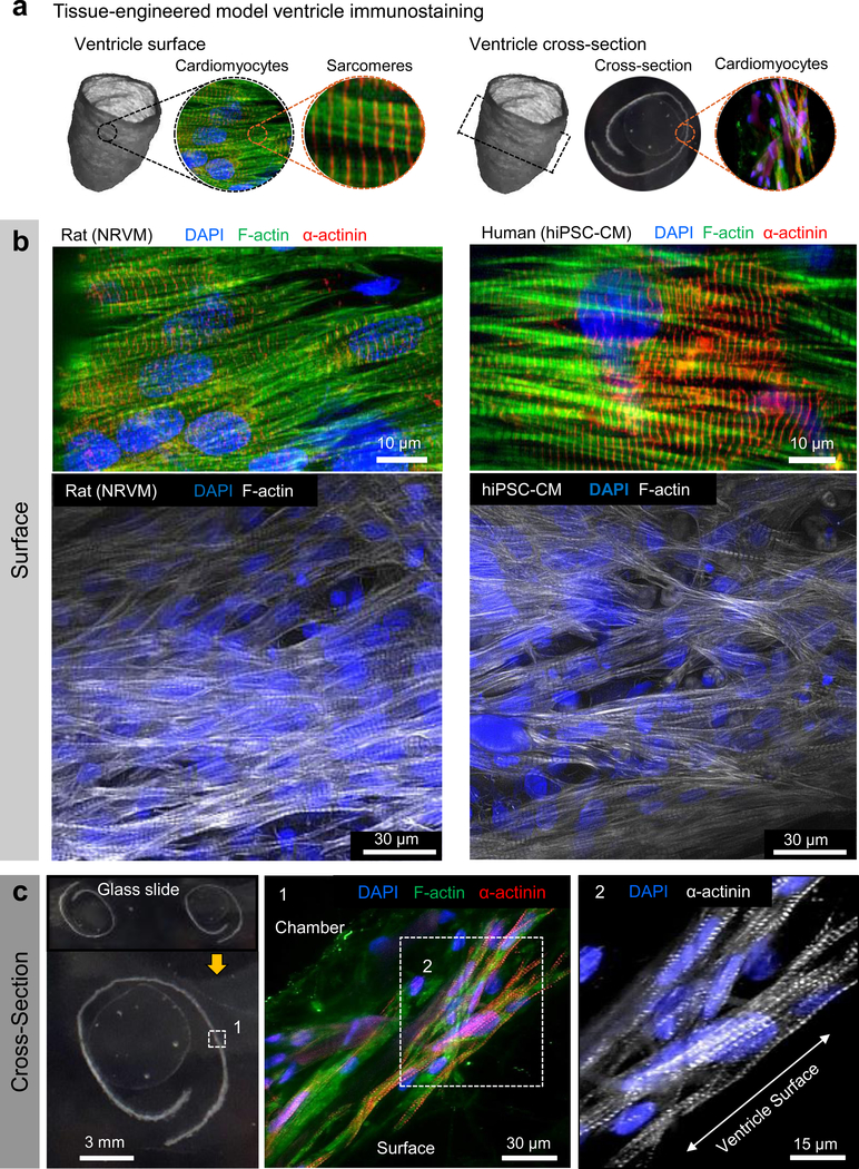
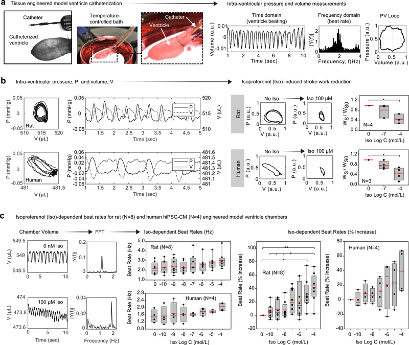
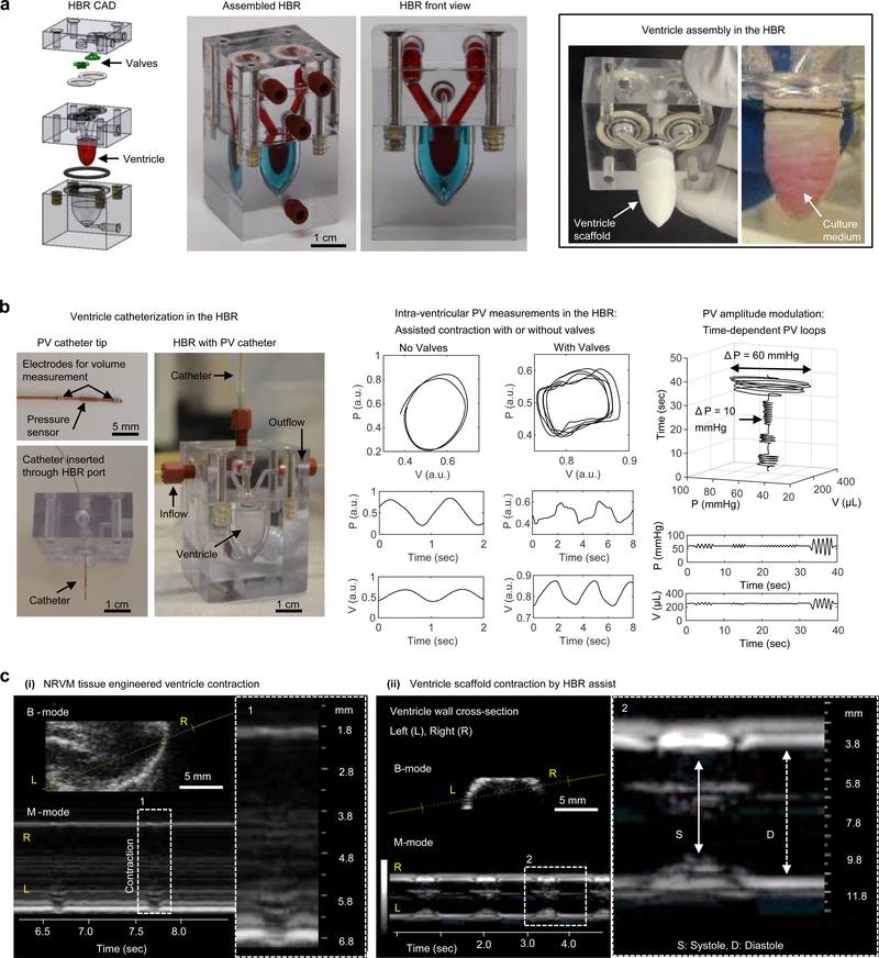
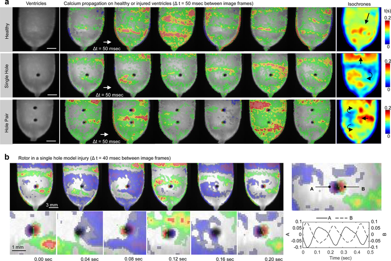
Laboratory studies of the heart use cell and tissue cultures to dissect heart function yet rely on animal models to measure pressure and volume dynamics. Here, we report tissue-engineered scale models of the human left ventricle, made of nanofibrous scaffolds that promote native-like anisotropic myocardial tissue genesis and chamber-level contractile function. Incorporating neonatal rat ventricular myocytes or cardiomyocytes derived from human induced pluripotent stem cells, the tissue-engineered ventricles have a diastolic chamber volume of ~500 µl (comparable to that of the native rat ventricle and approximately 1/250 the size of the human ventricle), and ejection fractions and contractile work 50-250 times smaller and 104-108 times smaller than the corresponding values for rodent and human ventricles, respectively. We also measured tissue coverage and alignment, calcium-transient propagation and pressure-volume loops in the presence or absence of test compounds. Moreover, we describe an instrumented bioreactor with ventricular-assist capabilities, and provide a proof-of-concept disease model of structural arrhythmia. The model ventricles can be evaluated with the same assays used in animal models and in clinical settings.


Conflict of interest statement

Competing interests

The authors declare no competing interests.

Figures

Figure 1|
Figure 1|. Tissue-engineered model ventricles recapitulate key structural and functional aspects of natural ventricular myocardium.
a, A schematic overview of the ventricle design-build-test project phases, from left to right: Human left ventricle ellipsoidal shape and fibrous extra-cellular matrix (ECM) inspired our use of circumferentially oriented nanofibers in scale-model ellipsoidal ventricle scaffolds. We seeded these scaffolds with cardiomyocytes to produce tissue-engineered ventricles and evaluated their contractile strength by pressure-volume catheterization. b, Pull spinning a nanofibrous ellipsoidal model ventricle scaffold: A high-speed rotating bristle dips into a fixed, continuous polymer source that is fed via a syringe pump through a small orifice (in this case a needle). The bristle pulls the polymer column into a polymer jet that is ejected towards a rotating collector. Solvent evaporation occurs as the polymer jet travels towards the collection mandrel, where the resulting nanofibers are collected. The resulting ventricle scaffolds are removed from the collector by tweezers in a hydration bath. c, Micro-computed tomography (μCT) images of a ventricle scaffold. d, Calcium propagation imaging performed on day 14. Spontaneous activity produced propagating waves but they did not always originate at the apex (left). Field stimulation, resulting from electrical pacing where electrodes were placed far from the ventricle surface, produced uniform calcium activation (middle). Apical point stimulation (indicated by yellow arrows) produced calcium waves that propagated from apex to base at a rate of 9.33 cm/sec for neonatal rat ventricular myocyte (NRVM) ventricles and 5.2 cm/sec for human induced pluripotent stem cell-derived cardiomyocyte (hiPSC-CM) ventricles (right). Calcium fluorescence intensity is displayed as a heat map ranging from blue (min) to red (max), overlaid on a greyscale image of the ventricle surface
Figure 2|
Figure 2|. Tissue-engineered ventricle immunostaining.
a, A schematic of ventricle surface or cross-section immunostains shown in b (surface) and c (cross-section). In all cases, cells were introduced via the exterior surface and imaging was performed following 14 days in culture. Immunostaining confirmed that both neonatal rat ventricular myocytes (NRVM) and human induced pluripotent stem cell-derived cardiomyocytes (Cor.4U hiPSC-CM) infiltrated the scaffolds and were aligned roughly circumferentially, coincident with the scaffold’s nanofiber ultrastructure.
Figure 3|
Figure 3|. Intra-ventricular pressure-volume (PV) data obtained by tissue-engineered ventricle catheterization.
a, An overview of the ventricle catheterization procedure. Catheters were fed through tubing on which the ventricle base was sutured. Ventricles were submerged within a 3.5 cm petri dish bath, which was mounted on a temperature-controlled heating stage. Catheter readouts fed to signal amplification instruments provided real-time measurements of intra-ventricular pressure and volume. b, Intra-ventricular pressure, P, and volume, V, measured by catheterization of neonatal rat ventricular myocyte (NRVM)- or human induced pluripotent stem cell-derived cardiomyocyte (Cor.4U hiPSC-CM)-based ventricles. Exposure to isoproterenol (Iso) reduced stroke work of both rat and human ventricles. Here, P and V were normalized by polynomial fit to remove measurement drift occurring over the course of multiple Iso doses. P-values were P=0.04 (NRVM, N=4 ventricles) and P=0.038 (Cor.4U, N=3 ventricles). c, Iso-dependent beat rates for rat (N=8 ventricles) and human (N=4 ventricles). Time-domain recordings of chamber volume were Fourier-transformed to obtain beat rates. The spontaneous beat rate of NRVM ventricles (~130 ±15 bpm) was higher than hiPSC-CM ventricles (~85 ± 15 bpm), and both increased by ~40% following exposure to 10−4 M Iso. * P < 0.05, ** P < 0.001, compared to baseline (no Iso), one-way ANOVA with Tukey post-hoc test. Exact P-values for Iso-dependent beat rates (Hz) were P=0.0325 (NRVM, N=8 ventricles) and P=0.524 (Cor.4U, N=4 ventricles). Exact P-values for Iso-dependent beat rate (% increase) were P<0.001 for baseline NRVM versus the highest Iso dose of 10−4 M. Exact P-values for NRVM at the smallest three doses (10−10 M, 10−9 M, and 10−8 M Iso) versus the largest dose (10−4 M Iso) were P=0.003, P=0.006, and P=0.008, respectively. The exact P-value for NRVM dosed with 10−9 M Iso versus 10−4 M Iso was P=0.011. For Cor.4U, differences in beat rate were not statistically significant (P=0.183). In all cases, measurements were performed on day 14. Data are presented as box plots with individual data points overlaid, where lower or upper edges of the box represent 25th or 75th percentiles, the middle bar is the median, dashed red bar is the mean, and whiskers are minimum and maximum values.
Figure 4|
Figure 4|. A heart bioreactor (HBR) for tissue-engineered ventricle culture, assisted contraction, and instrumentation.
a, An overview of ventricle and valve (optional) assembly within the HBR. A computer aided design (CAD) drawing of HBR components shows valve and ventricle placement. The ventricle wall separates intra- and extra- ventricular flow loops, which are indicated by red and blue dyes in the assembled HBR. Ventricle scaffolds are sutured over a support ring where input and output channels of the intra-ventricular flow loop converge. Pressure supplied by an external source to the extra-ventricular flow loop drives assisted ventricle contraction and flow through the intra-ventricular flow loop. b, Ventricle catheterization in the HBR enabled pressure and volume measurements during assisted ventricle contraction with or without cast-molded silicone tricuspid valves. c, Echocardiographic measurement of a tissue-engineered neonatal rat ventricular myocyte (NRVM, day 14) ventricle contracting unassisted (left) and a cell-free ventricle scaffold contracting by HBR assist (right). In both cases, cross-sections (brightness mode) and time-dependent traces of the ventricle wall (motion, M-mode) are shown.
Figure 5|
Figure 5|. Structural arrhythmia disease model.
a, Calcium wavefront propagation on a healthy (top) or injured (bottom) tissue engineered neonatal rat ventricular myocyte (NRVM) ventricle. The uninjured ventricle exhibited plane waves with a peak-to-peak spatial period of ~5 mm (top). Subsequent injury of this ventricle using a 1 mm diameter biopsy punch resulted in circular anatomical defects that generated pinned spiral waves. The single-hole injury generated a single spiral wave whereas the hole-pair generated counter-propagating spiral waves that converged and propagated through the inter-hole region with each cycle. b, Calcium fluorescence intensity measurements near the rotor poles of the single hole injury showed consistent phase difference and a rotation rate of ~ 5 Hz. Experiments were performed at day 12 and spontaneous activity was recorded without external stimulation for all cases. Measurements shown in (a) were acquired using a 5 msec exposure window, whereas those shown in (b) were acquired using a 10 msec exposure window. In all cases, the temporal derivative of calcium fluorescence intensity is displayed as a heat map ranging from blue (min) to red (max), overlaid on a greyscale image of the ventricle surface.

Comment in

References

    1. Benam KH et al. Engineered in vitro disease models. Annu Rev Pathol 10, 195–262, doi: 10.1146/annurev-pathol-012414-040418 (2015). - DOI - PubMed
    1. Tzatzalos E, Abilez OJ, Shukla P & Wu JC Engineered heart tissues and induced pluripotent stem cells: Macro- and microstructures for disease modeling, drug screening, and translational studies. Adv Drug Deliv Rev 96, 234–244, doi: 10.1016/j.addr.2015.09.010 (2016). - DOI - PMC - PubMed
    1. Pacher P, Nagayama T, Mukhopadhyay P, Batkai S & Kass DA Measurement of cardiac function using pressure-volume conductance catheter technique in mice and rats. Nat Protoc 3, 1422–1434, doi: 10.1038/nprot.2008.138 (2008). - DOI - PMC - PubMed
    1. Ram R, Mickelsen DM, Theodoropoulos C & Blaxall BC New approaches in small animal echocardiography: imaging the sounds of silence. American journal of physiology. Heart and circulatory physiology 301, H1765–1780, doi: 10.1152/ajpheart.00559.2011 (2011). - DOI - PMC - PubMed
    1. Bakermans AJ et al. Small animal cardiovascular MR imaging and spectroscopy. Progress in nuclear magnetic resonance spectroscopy 88-89, 1–47, doi: 10.1016/j.pnmrs.2015.03.001 (2015). - DOI - PubMed

Publication types