. 2019 Apr 16:19:101.
doi: 10.1186/s12935-019-0813-2. eCollection 2019.

Overexpression of HHLA2 in human clear cell renal cell carcinoma is significantly associated with poor survival of the patients

Affiliations

Overexpression of HHLA2 in human clear cell renal cell carcinoma is significantly associated with poor survival of the patients

Lujun Chen et al. Cancer Cell Int. .

Abstract

Background: It is well known that human clear cell renal cell carcinoma (ccRCC) is a highly immunogenic and chemo-resistant tumor. Recently, emerging data suggest that the immune checkpoint blockade therapy is an important breakthrough in the treatment against ccRCC. HHLA2, a recently reported member of B7 family, is uniquely expressed in humans but not in mice, and it plays an important role in the functional inhibition of CD4 and CD8 T cells. Herein, we aimed to study the clinical implications of HHLA2 expression in human ccRCC and its potential regulatory role in the biological functions of the cancer cells.

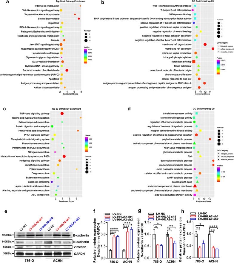
Methods: In the present study, we examined HHLA2 expression in human ccRCC tissues and analyzed the clinical implications as well as prognostic value. The intervention of HHLA2 in human ccRCC cell lines ACHN and 786-O was performed and its effect on the cellular function of the cells was also analyzed. We also identified the differentially expressed genes upon HHLA2 knockdown in ccRCC cell lines by using gene microarray analysis.

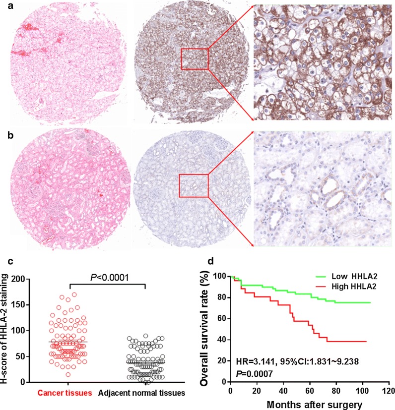
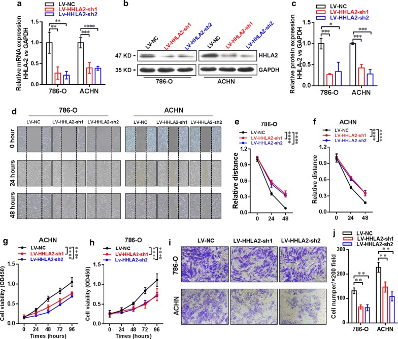
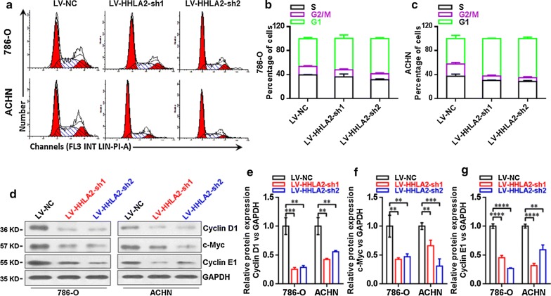
Results: We found that higher HHLA2 mRNA expression level in human ccRCC tissues compared with that in adjacent normal tissues based on TCGA data, and the HHLA2 expression at mRNA level was positively and significantly correlated with PD-L1, PD-L2, B7-H6, but negatively and significantly correlated with B7-H3. Moreover, our immunohistochemistry study showed that the staining intensity of HHLA2 in human ccRCC tissues was significantly higher than that in the adjacent normal tissues, and the overall survival rate of ccRCC patients with higher HHLA2 expression was significantly poorer than that of the patients with lower HHLA2 expression. Higher expression of HHLA2 in ccRCC tissues was positively and significantly associated with larger tumor size and advanced TNM stage. The COX model revealed that the parameters including patient's age, TNM stage and HHLA2 expression level could be used as the independent risk factors respectively for the prognostic prediction of the patients. Our cellular study showed that upon knockdown of HHLA2 expression in human ccRCC cell lines, the cell viability, the migration and the invasion ability were significantly inhibited, while the cell cycle arrest at G1 phase was induced and the expressions of Cyclin D1, c-Myc and Cyclin E1 were decreased. In addition, according to the microarray data, the expressions of epithelia-to-mesenchymal transition markers, such as E-cadherin, N-cadherin and Vimentin, were significantly changed after knockdown of HHLA2 expression.

Conclusions: Our findings indicated that HHLA2 was involved in the progression of human ccRCC and could be used as an important prognostic predictor for this malignancy.

Keywords: Clear cell renal cell carcinoma; Epithelial-to-mesenchymal transition; HHLA2; Prognosis.


Conflict of interest statement

The authors declare that they have no competing interests.

Figures

Fig. 1
Fig. 1
Survey of HHLA2 expression at mRNA level in human ccRCC tissues based on TCGA data. We compared the HHLA2 expression at mRNA level between human ccRCC tissues and adjacent normal tissues according to TCGA data from http://gepia.cancer-pku.cn/. a Higher expression in human ccRCC tissues was found compared with adjacent normal tissues (P < 0.05). The HHLA2 mRNA expression level was positively and significantly correlated with PD-L1 (b, P < 0.05), PD-L2 (c, P < 0.001), B7-H6 (f, P < 0.0001), but negatively and significantly correlated with B7-H3 (d, P < 0.05), and there was no significant association between HHLA2 and B7-H4 (e, P > 0.05)
Fig. 2
Fig. 2
HHLA2 expression in human ccRCC tissues. a The positive staining of HHLA2 could be predominantly found in the cytoplasm and on the membrane of the cancer cells in ccRCC tissues. b Weak staining of HHLA2 was found in adjacent normal tissues. c The staining intensity of HHLA2 in human ccRCC tissues was significantly higher than that in adjacent normal tissues (P < 0.0001). d The overall survival rate of ccRCC patients with low HHLA2 expression was significantly higher than that of the patients with high HHLA2 expression (HR = 3.141, 95% CI 1.831–9.238, P = 0.007)
Fig. 3
Fig. 3
Establishment of stable knockdown of HHLA2 expression in ccRCC cell lines 786-O and ACHN. a The knockdown efficiency was confirmed by real-time RT-PCR analysis, and the data showed that the mRNA expression level of HHLA2 was significantly decreased in the LV-HHLA2-sh1 (P < 0.01 in 786-O cells, and P < 0.001 in ACHN cells) and LV-HHLA2-sh2 (P < 0.01 in 786-O cells, and P < 0.0001 in ACHN cells) compared with LV-NC group cells respectively. b, c By using the western blotting analysis, we also confirmed that the HHLA2 expression at the protein level was significantly decreased in the LV-HHLA2-sh1 (P < 0.001 in 786-O cells, and P < 0.001 in ACHN cells) and LV-HHLA2-sh2 (P < 0.05 in 786-O cells, and P < 0.001 in ACHN cells) compared with LV-NC group cells. df The migration ability of the ccRCC cell lines after knockdown HHLA2 expression in the LV-HHLA2-sh1 and LV-HHLA2-sh2 groups was examined compared with the LV-NC group cells. At the time point of 48 h, the relative distance of LV-HHLA2-sh1 (P < 0.0001 in 786-O cells, and P < 0.0001 in ACHN cells) and LV-HHLA2-sh2 (P < 0.0001 in 786-O cells, and P < 0.0001 in ACHN cells) was significantly increased compared with the LV-NC group cells. g, h Our CCK-8 assay showed that the cell proliferation ability of ccRCC cell lines was significantly decreased in the LV-HHLA2-sh1 (P < 0.0001 in 786-O cells, and P < 0.0001 in ACHN cells) and LV-HHLA2-sh2 (P < 0.0001 in 786-O cells, and P < 0.0001 in ACHN cells) compared with the LV-NC group cells. i, j Transwell assay showed that the cell invasive ability of ccRCC cell lines was significantly decreased in the LV-HHLA2-sh1 (P < 0.01 in 786-O cells, and P < 0.01 in ACHN cells) and LV-HHLA2-sh2 (P < 0.01 in 786-O cells, and P < 0.01 in ACHN cells) compared with the LV-NC group cells
Fig. 4
Fig. 4
Expressions of Cyclin D1, c-Myc and Cyclin E1 in ccRCC cell lines after knockdown of HHLA2. ac The knockdown HHLA2 expression significantly increased the ratio of G1 phase and induced cell cycle arrest in human ccRCC cell lines. d Western blotting analysis of Cyclin D1, c-Myc and Cyclin E1 in ccRCC cell lines after knockdown of HHLA2 expression in 786-O and ACHN cells respectively. e The Cyclin D1 was significantly decreased in the LV-HHLA2-sh1 (P < 0.01 in 786-O cells, and P < 0.01 in ACHN cells) and LV-HHLA2-sh2 (P < 0.01 in 786-O cells, and P < 0.01 in ACHN cells) compared with the LV-NC group cells. f The c-Myc was significantly decreased in the LV-HHLA2-sh1 (P < 0.01 in 786-O cells, and P < 0.01 in ACHN cells) and LV-HHLA2-sh2 (P < 0.01 in 786-O cells, and P < 0.001 in ACHN cells) compared with the LV-NC group cells. g The Cyclin E1 was significantly decreased in the LV-HHLA2-sh1 (P < 0.0001 in 786-O cells, and P < 0.0001 in ACHN cells) and LV-HHLA2-sh2 (P < 0.0001 in 786-O cells, and P < 0.01 in ACHN cells) compared with the LV-NC group cells
Fig. 5
Fig. 5
HHLA2 is potentially involved in the regulation of EMT in human ccRCC cells. We identified the differentially expressed gene profiles between LV-HHLA2-sh1 and LV-NC groups of ccRCC cell lines using microarray analysis. a, b The co-up-regulated genes were involved in the KEGG and GO analyses, and the top 20 pathways were listed, and for the co-down-regulated genes, the top 20 pathways were listed in c, d. In the following cellular study, we further examined the changes of EMT markers after HHLA2 knockdown expression in ccRCC cell lines. e Western blotting analysis of E-cadherin, N-cadherin and Vimentin in 786-O and ACHN cells in different groups. f After knockdown of HHLA2 in 786-O and ACHN, the expression of E-cadherin was significantly increased (In 786-O: LV-HHLA2-sh1 vs LV-NC: P < 0.001, LV-HHLA2-sh2 vs LV-NC: P < 0.0001; In ACHN: LV-HHLA2-sh1 vs LV-NC: P < 0.001, LV-HHLA2-sh2 vs LV-NC: P < 0.001). g After knockdown of HHLA2 in 786-O and ACHN, the expression of N-cadherin was significantly decreased (In 786-O: LV-HHLA2-sh1 vs LV-NC: P < 0.05, LV-HHLA2-sh2 vs LV-NC: P < 0.05; In ACHN: LV-HHLA2-sh1 vs LV-NC: P < 0.01, LV-HHLA2-sh2 vs LV-NC: P < 0.05). h After knockdown of HHLA2 in 786-O and ACHN, the expression of Vimentin was significantly decreased (In 786-O: LV-HHLA2-sh1 vs LV-NC: P < 0.01, LV-HHLA2-sh2 vs LV-NC: P < 0.05; In ACHN: LV-HHLA2-sh1 vs LV-NC: P < 0.001, LV-HHLA2-sh2 vs LV-NC: P < 0.0001)

References

    1. Siegel RL, Miller KD, Jemal A. Cancer statistics, 2018. CA Cancer J Clin. 2018;68(1):7–30. doi: 10.3322/caac.21442. - DOI - PubMed
    1. Rini BI, Campbell SC, Escudier B. Renal cell carcinoma. Lancet. 2009;373(9669):1119–1132. doi: 10.1016/S0140-6736(09)60229-4. - DOI - PubMed
    1. Lopez-Beltran A, Scarpelli M, Montironi R, Kirkali Z. 2004 WHO classification of the renal tumors of the adults. Eur Urol. 2006;49(5):798–805. doi: 10.1016/j.eururo.2005.11.035. - DOI - PubMed
    1. Rini BI, Rathmell WK, Godley P. Renal cell carcinoma. Curr Opin Oncol. 2008;20(3):300–306. doi: 10.1097/CCO.0b013e3282f9782b. - DOI - PubMed
    1. Mogami T, Harada J, Kishimoto K, Sumida S. Percutaneous MR-guided cryoablation for malignancies, with a focus on renal cell carcinoma. Int J Clin Oncol. 2007;12(2):79–84. doi: 10.1007/s10147-006-0654-6. - DOI - PubMed