. 2019 Apr 16:4:8.
doi: 10.1038/s41536-019-0070-y. eCollection 2019.

Proteomic analysis of human mesenchymal stromal cell secretomes: a systematic comparison of the angiogenic potential

Affiliations

Proteomic analysis of human mesenchymal stromal cell secretomes: a systematic comparison of the angiogenic potential

Debora Kehl et al. NPJ Regen Med. .

Abstract

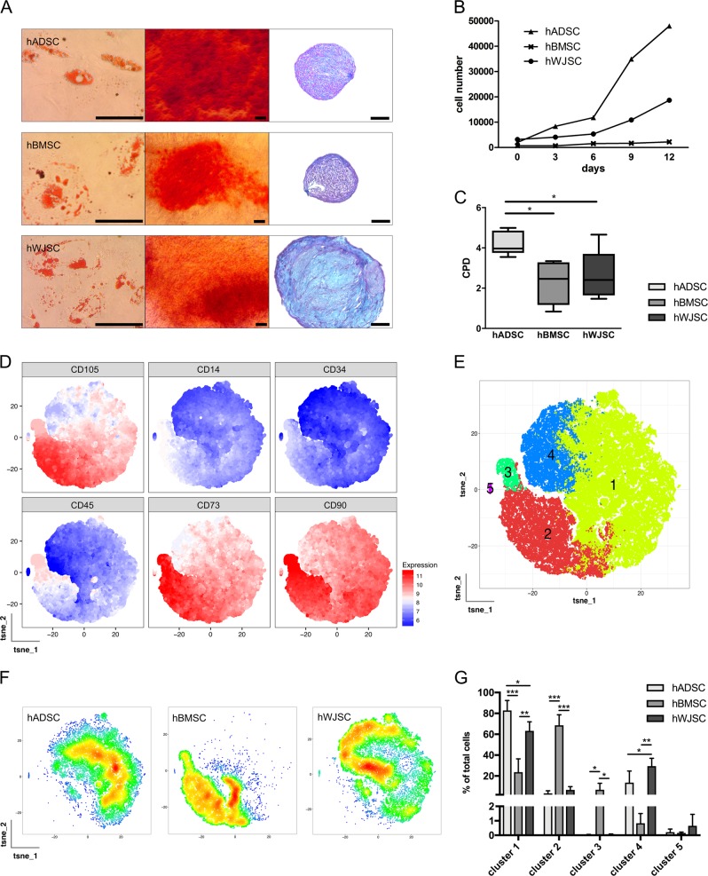
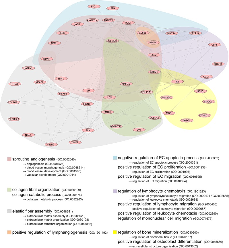
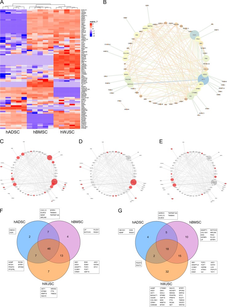
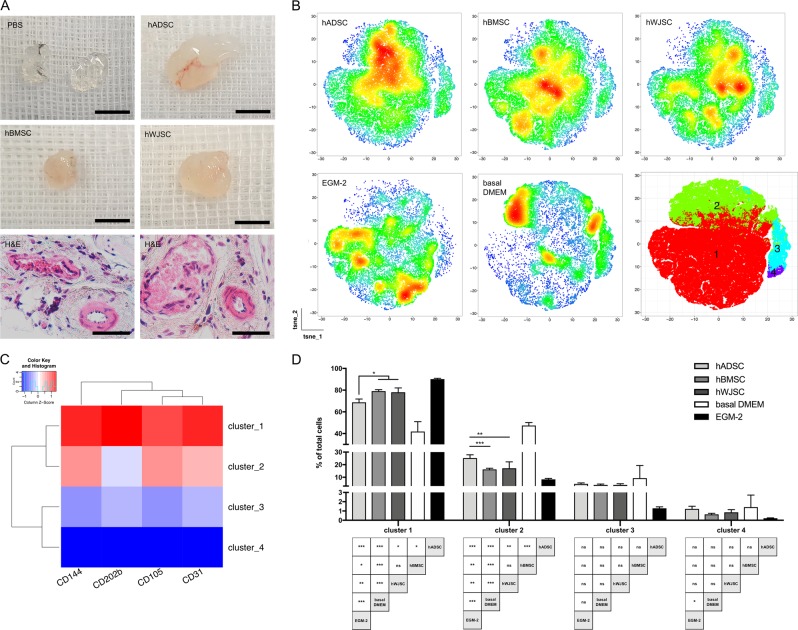
Human mesenchymal stromal cell (hMSC) secretomes have shown to influence the microenvironment upon injury, promoting cytoprotection, angiogenesis, and tissue repair. The angiogenic potential is of particular interest for the treatment of ischemic diseases. Interestingly, hMSC secretomes isolated from different tissue sources have shown dissimilarities with respect to their angiogenic profile. This study compares angiogenesis of hMSC secretomes from adipose tissue (hADSCs), bone marrow (hBMSCs), and umbilical cord Wharton's jelly (hWJSCs). hMSC secretomes were obtained under xenofree conditions and analyzed by liquid chromatography tandem mass spectrometry (LC/MS-MS). Biological processes related to angiogenesis were found to be enriched in the proteomic profile of hMSC secretomes. hWJSC secretomes revealed a more complete angiogenic network with higher concentrations of angiogenesis related proteins, followed by hBMSC secretomes. hADSC secretomes lacked central angiogenic proteins and expressed most detected proteins to a significantly lower level. In vivo all secretomes induced vascularization of subcutaneously implanted Matrigel plugs in mice. Differences in secretome composition were functionally analyzed with monocyte and endothelial cell (EC) in vitro co-culture experiments using vi-SNE based multidimensional flow cytometry data analysis. Functional responses between hBMSC and hWJSC secretomes were comparable, with significantly higher migration of CD14++ CD16- monocytes and enhanced macrophage differentiation compared with hADSC secretomes. Both secretomes also induced a more profound pro-angiogenic phenotype of ECs. These results suggest hWJSCs secretome as the most potent hMSC source for inflammation-mediated angiogenesis induction, while the potency of hADSC secretomes was lowest. This systematic analysis may have implication on the selection of hMSCs for future clinical studies.


Conflict of interest statement

The authors declare no competing interests.

Figures

Fig. 1
Fig. 1
Characterization of hMSC from adipose tissue, bone marrow and umbilical cord Wharton’s jelly. a Trilineage differentiation of hMSCs (n = 5 per hMSC tissue source) into adipogenic (left; Oil Red O staining), osteogenic (middle; Alzarin Red S) and chondrogenic (right; Alcian Blue PAS) lineages. Scale bar = 100 μm. b Proliferation capacities of hMSCs (n = 5 per hMSC tissue source) over 12 days, c showing the cumulative population doublings of each hMSC source. d All hMSCs (n = 5 per hMSC tissue source) positively expressed CD73, CD90, CD105, and only a minimal proportion of cells were positive for CD14, CD34 and CD45. Data are visualized by bi-dimensional t-SNE maps generated from multidimensional flow cytometry data. e Recorded events were subdivided into 5 clusters by PhenoGraph and FlowSom algorithms according to surface marker expression. The cellular distribution of 5 donors per cell source is f visualized with cumulative density plots and g quantified accordingly. Bar graphs present mean ± s.d. (*p < 0.05, **p < 0.01, ***p < 0.001; one-way ANOVA and Tukey multiple comparison)
Fig. 2
Fig. 2
LC/MS-MS analysis of hMSC CM. a Heat maps (red = expressed, blue = not expressed) and b Venn diagrams represent the total amount of identified proteins in hMSC CM (n = 5 per hMSC tissue source), where more exclusive proteins and higher protein intensities were found for hWJSC CM. Identified proteins found in all hMSC CM were characterized on their d cellular location and e function
Fig. 3
Fig. 3
Enriched biological processes in hMSC CM. LC/MS-MS data were analyzed on overrepresented biological processes in the extracellular protein fraction of hMSC CM (n = 5 per hMSC tissue source). The most relevant biological processes, their protein-protein interactions and corresponding coloring patterns are displayed
Fig. 4
Fig. 4
The pro-angiogenic proteome of hMSC CM. a Heat map generated based on protein intensities displays hMSC CM proteins involved in angiogenesis (n = 5 per hMSC tissue source; red = expressed, blue = not expressed). b Interactions of angiogenesis related proteins, where node size/color depends on the amount of protein interactions. Proteins not found in the according CM are displayed in red, demonstrating that central pro-angiogenic proteins are missing in c hADSC CM compared to d hBMSC CM and e hWJSC CM. f The protein composition of hMSC CM and their exclusive proteins demonstrate that hBMSC CM and hWJSC CM are closer related compared to hADSC CM; g however significantly higher protein intensities by pairwise student t-test were found for hWJSC CM
Fig. 5
Fig. 5
Immune cell response to hMSC CM. a hMSC CM (n = 5 per tissue source) revealed enhanced migration of monocytes (n = 3 healthy blood donors), with significantly higher migration induced by hBMSC CM and hWJSC CM in comparison to hADSC CM and 5 ng/ml MCP-1 control. b Migrated monocytes were characterized by CD14 and CD16 staining, c demonstrating that hMSC CM mainly induced migration of classical CD14++ CD16 monocytes, but not of intermediate and non-classical monocytes. d 7 myeloid cell populations were identified after co-incubation of monocytic Thp-1 cells and hMSC CM (n = 5 per tissue source) displayed by a bi-dimensional cumulative vi-SNE map. e The percentages of each cell population were monitored, including f classical monocytes, g macrophages, and h all (diff.) (plasmacytoid) dendritic cells. hBMSC CM and hWJSC CM led to a significantly higher monocyte to macrophage differentiation in comparison to hADSC CM and basal DMEM control. Bar graphs present mean ± s.d. (*p < 0.05, **p < 0.01, ***p < 0.001; one-way ANOVA and Tukey multiple comparison)
Fig. 6
Fig. 6
hMSC secretome is pro-angiogenic in vitro and in vivo. a In vivo Matrigel plug assay demonstrated significantly increased vascularization when using hMSC CM compared to basal medium only. Scale bar = 0.5 cm / H&E = 100 μm. bd Stimulation of HUVEC with hMSC CM (n = 5 per tissue source) induces expression of angiogenesis related markers, including CD31, CD105, CD202b, and CD144. b The density distribution of cells was dependent on the co-incubation medium. Clustering flow cytometry data defined 4 different clusters, c each with a distinct expression profile. d A significantly higher shift towards the pro-angiogenic cluster 1 was found when HUVEC were co-incubated with hBMSC CM and hWJSC CM. Bar graphs present mean ± s.d. (*p < 0.05, **p < 0.01, ***p < 0.001; one-way ANOVA and Tukey multiple comparison)

Similar articles

Cited by

References

    1. Galipeau J, Sensebe L. Mesenchymal stromal cells: clinical challenges and therapeutic opportunities. Cell. Stem. Cell. 2018;22:824–833. doi: 10.1016/j.stem.2018.05.004. - DOI - PMC - PubMed
    1. Baraniak PR, McDevitt TC. Stem cell paracrine actions and tissue regeneration. Regen. Med. 2010;5:121–143. doi: 10.2217/rme.09.74. - DOI - PMC - PubMed
    1. Murphy MB, Moncivais K, Caplan AI. Mesenchymal stem cells: environmentally responsive therapeutics for regenerative medicine. Exp. Mol. Med. 2013;45:e54. doi: 10.1038/emm.2013.94. - DOI - PMC - PubMed
    1. Tao H, Han Z, Han ZC, Li Z. Proangiogenic features of mesenchymal stem cells and their therapeutic applications. Stem Cells Int. 2016;2016:1314709. doi: 10.1155/2016/1314709. - DOI - PMC - PubMed
    1. Kwon HM, et al. Multiple paracrine factors secreted by mesenchymal stem cells contribute to angiogenesis. Vasc. Pharmacol. 2014;63:19–28. doi: 10.1016/j.vph.2014.06.004. - DOI - PubMed