. 2019 Feb 10;6(8):1801233.
doi: 10.1002/advs.201801233. eCollection 2019 Apr 17.

Inhibition of CaMKIIα Activity Enhances Antitumor Effect of Fullerene C60 Nanocrystals by Suppression of Autophagic Degradation

Affiliations

Inhibition of CaMKIIα Activity Enhances Antitumor Effect of Fullerene C60 Nanocrystals by Suppression of Autophagic Degradation

Jing Xu et al. Adv Sci (Weinh). .

Abstract

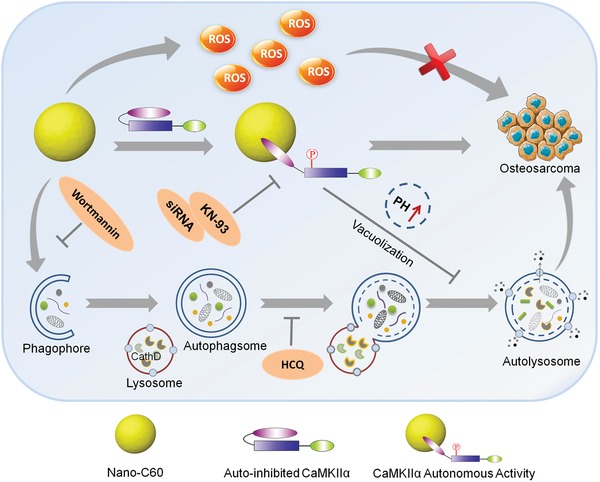
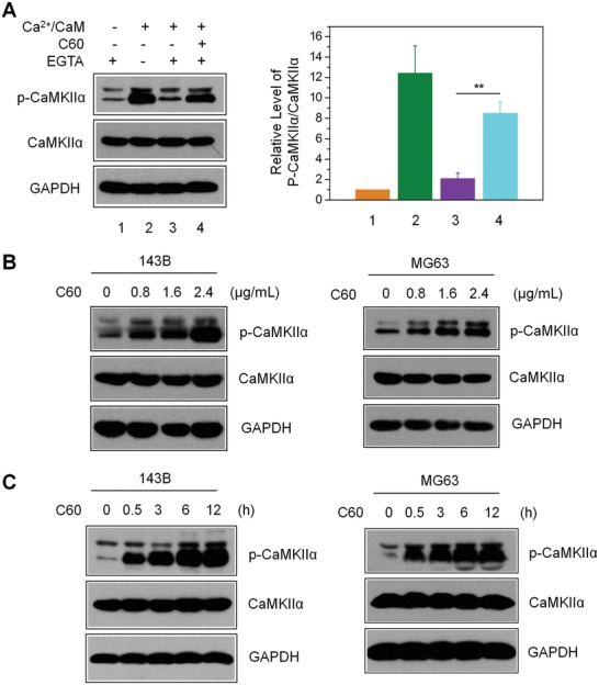
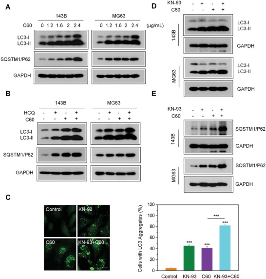
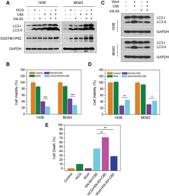
Fullerene C60 nanocrystals (nano-C60) possess various attractive bioactivities, including autophagy induction and calcium/calmodulin-dependent protein kinase IIα (CaMKIIα) activation. CaMKIIα is a multifunctional protein kinase involved in many cellular processes including tumor progression; however, the biological effects of CaMKIIα activity modulated by nano-C60 in tumors have not been reported, and the relationship between CaMKIIα activity and autophagic degradation remains unclear. Herein, nano-C60 is demonstrated to elicit reactive oxygen species (ROS)-dependent cytotoxicity and persistent activation of CaMKIIα in osteosarcoma (OS) cells. CaMKIIα activation, in turn, produces a protective effect against cytotoxicity from nano-C60 itself. Inhibition of CaMKIIα activity by either the chemical inhibitor KN-93 or CaMKIIα knockdown dramatically promotes the anti-OS effect of nano-C60. Moreover, inhibition of CaMKIIα activity causes lysosomal alkalinization and enlargement, and impairs the degradation function of lysosomes, leading to autophagosome accumulation. Importantly, excessive autophagosome accumulation and autophagic degradation blocking are shown to play an important role in KN-93-enhanced-OS cell death. The synergistic anti-OS efficacy of KN-93 and nano-C60 is further revealed in an OS-xenografted murine model. The results demonstrate that CaMKIIα inhibition, along with the suppression of autophagic degradation, presents a promising strategy for improving the antitumor efficacy of nano-C60.

Keywords: autophagic degradation; autophagy; calcium/calmodulin‐dependent protein kinase IIα; fullerene C60 nanocrystals; osteosarcoma therapy.


Conflict of interest statement

The authors declare no conflict of interest.

Figures

Scheme 1
Scheme 1
Schematic illustration of combination treatment strategy of nano‐C60 and CaMKIIα inhibition for OS therapy.
Figure 1
Figure 1
Nano‐C60‐induced autonomous CaMKIIα activity in OS cells. A) T286 autophosphorylation assay for CaMKIIα in 143B cell lysates, as detected by anti‐CaMKII and phospho‐CaMKII antibodies. The right panel shows the level of p‐CaMKIIα relative to total CaMKIIα, with the value for control (without Ca2+/CaM and nano‐C60) set at 1. Mean ± SEM, n = 3. **P < 0.01. B) Dose‐dependent CaMKIIα‐T286 autophosphorylation level in 143B and MG63 cells treated with nano‐C60 for 12 h. C) Time course of CaMKIIα‐T286 autophosphorylation levels in 143B and MG63 cells treated with 2.4 µg mL−1 nano‐C60.
Figure 2
Figure 2
Effects of CaMKIIα inhibition on nano‐C60‐induced cytotoxicity in OS cells. A)143B and MG63 cells were treated with 1.6 µg/mL−1 of nano‐C60 in the presence or absence of 10.0 × 10⁻⁶ m KN‐93 for 24 h. CaMKIIα level was detected by Western blotting with antibodies against CaMKII and phospho‐CaMKII. The right panel demonstrates the level of p‐CaMKIIα relative to that of total CaMKIIα, with the control value (without nano‐C60) set at 1. Mean ± SEM, n = 3. *P < 0.05, **P < 0.01. B) 143B cells were treated with or without 1.6 µg mL−1 of nano‐C60 in the presence or absence of 5.0 or 10.0 × 10⁻⁶ m KN‐93 for 24 h. Cell viability was measured by CCK‐8 assay. Mean ± SEM, n = 3. ***P < 0.005. C) Cell death assay of 143B cells treated as in A). Cell death rates were determined by Hoechst/PI staining and demonstrated as the percentage of PI‐positive cells. Mean ± SEM, n = 3. ***P < 0.005. D) Cell viability of 143B and MG63 cells treated with or without 1.6 µg mL−1 of nano‐C60 for 24 h after transfection with CaMKIIα siRNA or control siRNA for 48 h. Mean ± SEM, n = 3. **P < 0.01, ***P < 0.005. E) The cell death rates of 143B cells treated as described in D). Mean ± SEM, n = 3. ***P < 0.005.
Figure 3
Figure 3
Enhancement of nano‐C60‐induced incomplete autophagy by CaMKIIα inhibition. A) Western blotting for the expression of autophagy‐associated proteins in 143B and MG63 cells treated with different doses of nano‐C60 for 24 h using antibodies against LC3, SQATM1/P62, and GAPDH. B) 143B cells were treated with or without 1.6 µg mL−1 nano‐C60 in the presence or absence of 100.0 × 10⁻⁶ m HCQ for 24 h. LC3 and SQATM1/P62 levels were examined by Western blotting with anti‐LC3 and anti‐SQATM1/P62 antibodies, respectively. C) Representative fluorescence microscopy images of MG63‐EGFP‐LC3 cells treated with or without 1.6 µg mL−1 of nano‐C60 in the presence or absence of 10.0 × 10⁻⁶ m KN‐93 for 24 h. Scale bar = 20.0 × 10⁻⁶ m . The right panel demonstrates the rate of EGFP‐LC3 dots‐positive cells. Mean ± SEM, n = 3. ***P < 0.005. D,E) Western blot analysis of the expression of LC3‐II and SQATM1/P62 in 143B and MG63 cells treated with or without 1.6 µg mL−1 of nano‐C60 in the presence or absence of 10.0 × 10⁻⁶ m KN‐93 for 24 h.
Figure 4
Figure 4
Lysosomal alkalinization and enlargement caused by nano‐C60 and KN‐93 combination treatment. A) Representative fluorescence images of 143B‐RFP‐Lamp1 cells treated with or without 1.6 µg mL−1 of nano‐C60 in the presence or absence of 10.0 × 10⁻⁶ m KN‐93 for 16 h. Nuclei were stained with DAPI (blue). Scale bar = 20.0 µm. B) Representative fluorescence images of lysosome acidity using LysoSensor Green DND‐189 staining in 143B‐RFP‐Lamp1 cells treated with or without 1.0 µg mL−1 of nano‐C60 in the presence or absence of 7.5 × 10⁻⁶ m KN‐93 for 12 h. Scale bar = 50.0 µm. C) Flow cytometric analysis of lysosome acidity in 143B cells treated as described in B).
Figure 5
Figure 5
Impaired lysosomal degradation capacity caused by nano‐C60 and KN‐93 combination treatment. A) DQ‐BSA analysis of lysosomal proteolytic activity in 143B cells treated with or without 1.0 µg mL−1 of nano‐C60 in the presence or absence of 7.5 × 10⁻⁶ m KN‐93 for 12 h. The average number of the red fluorescent fragments in each cell was quantified (lower panel). At least 60 cells were analyzed for each treatment. Mean ± SEM, n = 3. **P < 0.01 ***P < 0.005. Scale bar = 50.0 µm. B) and C) Western blot analysis of enzymatic activity of cathepsin B and cathepsin D in 143B and MG63 cells treated with or without 1.6 µg mL−1 of nano‐C60 in the presence or absence of 10.0 × 10⁻⁶ m KN‐93 for 24 h.
Figure 6
Figure 6
Effects of autophagosome accumulation on cytotoxicity induced by nano‐C60 and KN‐93 combination treatment. A,B) Before 1.6 µg mL−1 of nano‐C60 and 10.0 × 10⁻⁶ m KN‐93 addition, 143B cells were pretreated with or without 100.0 × 10⁻⁶ m HCQ. A) LC3 and SQATM1/P62 levels were detected by Western blotting using anti‐LC3 and anti‐SQATM1/P62 antibodies, respectively, and B) cell viability was assessed by CCK‐8 assay. Mean ± SEM, n = 3. ***P < 0.005. C) Before 1.6 µg mL−1 of nano‐C60 and 10 × 10−6 m KN‐93 addition, 143B and MG63 cells were pretreated with or without 100.0 × 10⁻⁹ m Wort. LC3 levels were detected by Western blotting using anti‐LC3 and anti‐SQATM1/P62 antibodies, respectively. D) Cell viability of each group treated as described in C) was assessed by CCK‐8 assay. Mean ± SEM, n = 3. *P < 0.05, **P < 0.01. E) Before nano‐C60 and KN‐93 addition, 143B cells were pretreated with or without HCQ or Wort. Cell death assay was determined by Hoechst/PI staining and demonstrated as the percentage of PI‐positive cells. Mean ± SEM, n = 3. **P < 0.01. Scale bar = 200.0 µm.
Figure 7
Figure 7
Enhanced synergistic anti‐OS efficacy of nano‐C60 by CaMKIIα inhibitor KN‐93 in vivo. A) Tumors from nude mice treated with PBS (control), 0.5 mg kg−1 KN‐93 (s.c.), 0.2 mg kg−1 nano‐C60 (s.c.), or 0.5 mg kg−1 KN‐93 plus 0.2 mg kg−1 nano‐C60 (s.c.). B) Tumor volume and C) tumor weight in each group are shown. Mean ± SEM, n = 5. **P < 0.01, ***P < 0.005. D) TUNEL staining (green) of tumor tissues was performed to detect apoptotic cells, and nuclei were treated with DAPI (blue). Scale bar = 50.0 µm. E) Western blot analysis of the levels of phospho‐ or total‐CaMKIIα, and LC3 in tumor tissues.

Similar articles

Cited by

  • Guidelines for the use and interpretation of assays for monitoring autophagy (4th edition)1.
    Klionsky DJ, Abdel-Aziz AK, Abdelfatah S, Abdellatif M, Abdoli A, Abel S, Abeliovich H, Abildgaard MH, Abudu YP, Acevedo-Arozena A, Adamopoulos IE, Adeli K, Adolph TE, Adornetto A, Aflaki E, Agam G, Agarwal A, Aggarwal BB, Agnello M, Agostinis P, Agrewala JN, Agrotis A, Aguilar PV, Ahmad ST, Ahmed ZM, Ahumada-Castro U, Aits S, Aizawa S, Akkoc Y, Akoumianaki T, Akpinar HA, Al-Abd AM, Al-Akra L, Al-Gharaibeh A, Alaoui-Jamali MA, Alberti S, Alcocer-Gómez E, Alessandri C, Ali M, Alim Al-Bari MA, Aliwaini S, Alizadeh J, Almacellas E, Almasan A, Alonso A, Alonso GD, Altan-Bonnet N, Altieri DC, Álvarez ÉMC, Alves S, Alves da Costa C, Alzaharna MM, Amadio M, Amantini C, Amaral C, Ambrosio S, Amer AO, Ammanathan V, An Z, Andersen SU, Andrabi SA, Andrade-Silva M, Andres AM, Angelini S, Ann D, Anozie UC, Ansari MY, Antas P, Antebi A, Antón Z, Anwar T, Apetoh L, Apostolova N, Araki T, Araki Y, Arasaki K, Araújo WL, Araya J, Arden C, Arévalo MA, Arguelles S, Arias E, Arikkath J, Arimoto H, Ariosa AR, Armstrong-James D, Arnauné-Pelloquin L, Aroca A, Arroyo DS, Arsov I, Artero R, Asaro DML, Aschner M, Ashrafizadeh M, Ashur-Fabian O, Atanasov AG, Au AK, Auberger P, Auner HW, Aurelian L, Autelli R… See abstract for full author list ➔ Klionsky DJ, et al. Autophagy. 2021 Jan;17(1):1-382. doi: 10.1080/15548627.2020.1797280. Epub 2021 Feb 8. Autophagy. 2021. PMID: 33634751 Free PMC article.
  • Development of a novel autophagy-related gene model for gastric cancer prognostic prediction.
    Xu H, Xu B, Hu J, Xia J, Tong L, Zhang P, Yang L, Tang L, Chen S, Du J, Wang Y, Li Y. Xu H, et al. Front Oncol. 2022 Oct 7;12:1006278. doi: 10.3389/fonc.2022.1006278. eCollection 2022. Front Oncol. 2022. PMID: 36276067 Free PMC article.
  • miR-4295 promotes cell proliferation, migration and invasion of osteosarcoma through targeting interferon regulatory factor 1.
    Cheng JP, Huang B, Duan JH, Yi KJ, Zhuang ZL. Cheng JP, et al. Oncol Lett. 2020 Nov;20(5):260. doi: 10.3892/ol.2020.12123. Epub 2020 Sep 18. Oncol Lett. 2020. PMID: 32989394 Free PMC article.
  • Reductive damage induced autophagy inhibition for tumor therapy.
    Wang Y, Huang Y, Fu Y, Guo Z, Chen D, Cao F, Ye Q, Duan Q, Liu M, Wang N, Han D, Qu C, Tian Z, Qu Y, Zheng Y. Wang Y, et al. Nano Res. 2023;16(4):5226-5236. doi: 10.1007/s12274-022-5139-z. Epub 2022 Nov 22. Nano Res. 2023. PMID: 36465522 Free PMC article.
  • Nanotechnology for angiogenesis: opportunities and challenges.
    Kargozar S , Baino F , Hamzehlou S , Hamblin MR , Mozafari M . Kargozar S , et al. Chem Soc Rev. 2020 Jul 21;49(14):5008-5057. doi: 10.1039/c8cs01021h. Epub 2020 Jun 15. Chem Soc Rev. 2020. PMID: 32538379 Free PMC article. Review.

References

    1. Sun X., Huang X., Guo J., Zhu W., Ding Y., Niu G., Wang A., Kiesewetter D. O., Wang Z. L., Sun S., Chen X., J. Am. Chem. Soc. 2014, 136, 1706. - PMC - PubMed
    1. Zhao R., Han X., Li Y., Wang H., Ji T., Zhao Y., Nie G., ACS Nano 2017, 11, 8103. - PubMed
    1. Tong S., Quinto C. A., Zhang L., Mohindra P., Bao G., ACS Nano 2017, 11, 6808. - PubMed
    1. Riedinger A., Avellini T., Curcio A., Asti M., Xie Y., Tu R., Marras S., Lorenzoni A., Rubagotti S., Iori M., Capponi P. C., Versari A., Manna L., Seregni E., Pellegrino T., J. Am. Chem. Soc. 2015, 137, 15145. - PubMed
    1. Sherlock S. P., Tabakman S. M., Xie L., Dai H., ACS Nano 2011, 5, 1505. - PMC - PubMed

LinkOut - more resources