. 2019 Feb 25;6(8):1801735.
doi: 10.1002/advs.201801735. eCollection 2019 Apr 17.

Label-Free Multiphoton Endomicroscopy for Minimally Invasive In Vivo Imaging

Affiliations

Label-Free Multiphoton Endomicroscopy for Minimally Invasive In Vivo Imaging

Ashwathama Dilipkumar et al. Adv Sci (Weinh). .

Abstract

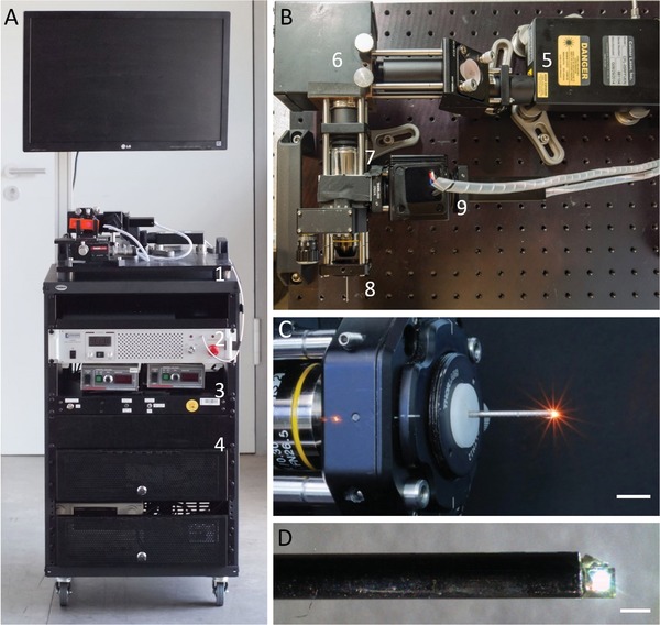
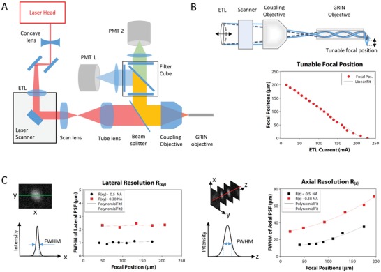
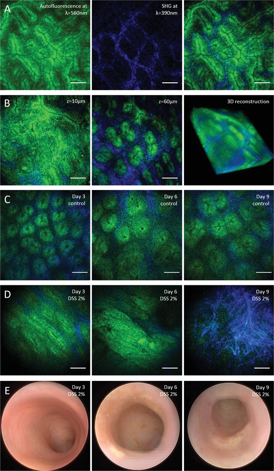
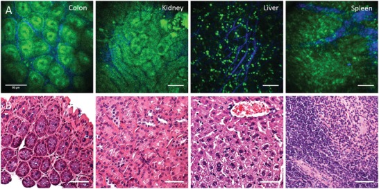
Multiphoton microscopy of cellular autofluorescence and second harmonic generation from collagen facilitates imaging of living cells and tissues without the need for additional fluorescent labels. Here, a compact multiphoton endomicroscope for label-free in vivo imaging in small animals via side-viewing needle objectives is presented. Minimal invasive imaging at cellular resolution is performed in colonoscopy of mice without surgical measures and without fluorescent dyes as a contrast agent. The colon mucosa is imaged repeatedly in the same animal in a mouse model of acute intestinal inflammation to study the process of inflammation at the tissue level within a time period of ten days, demonstrating the capabilities of label-free endomicroscopy for longitudinal studies for the first time.

Keywords: endoscopy; inflammatory bowel disease; multiphoton microscopy; optical imaging.


Conflict of interest statement

The authors declare no conflict of interest.

Figures

Figure 1
Figure 1
Multiphoton endomicroscope in a compact custom‐built setup. A) Transportable standalone system including (1) scanning microscope, (2) laser, (3) electronics, and (4) PC. B) Top view of the endomicroscope showing (5) laser head, (6) laser scanner, scan lens, and (7) tube lens, (8) coupling and endomicroscope objective, and (9) PMT detectors. C) Coupling objective and mounted endomicroscope objective. Scale bar: 1 cm. D) Endomicroscope objective with front‐mounted prism for side view. Scale bar: 1 mm. For in vivo experiments, the objective was mounted inside a 1.4 mm steel cannula and a droplet of glue was added to the back of the prism to cover its sharp edges and facilitate smooth positioning during colonoscopy.
Figure 2
Figure 2
Optical setup and performance. A) Schematic drawing of the optical path from laser head to endoscope objective and back to PMT detectors. See Experimental Section for further details. B) Illustration of focal plane shifting by means of an ETL and dependency of the focal position on the actuation current. C) Lateral and axial resolution measured as the size of the point spread function of 30 nm sized fluorescent beads dependent on the focal position.
Figure 3
Figure 3
Label‐free multiphoton endomicroscopy of murine colon mucosa in vivo. A) Two‐photon excited AF from endogenous molecules such as NADH or FAD (shown in green) and SHG from collagen‐I (shown in blue) show the epithelial crypt structure and underlying lamina of the colon mucosa. B) Focal plane shifting by means of a fast ETL for 3D visualization of the tissue morphology. C) Repeated minimal invasive multiphoton endomicroscopy of healthy murine colon as a control for the colitis model. D) Changes in the tissue morphology during the time course of intestinal inflammation in a mouse model of ulcerative colitis ranging from deformations of the crypt pattern at day 3, to massive epithelial erosion and collagen matrix formation at day 9. Images were recorded in the same animal with minimal invasive multiphoton endomicroscopy. E) Conventional wide‐field endoscopy as reference showing clear symptoms of acute murine DSS colitis from day 6 to 9, and macroscopically unchanged mucosa at day 3. Scale bars: 50 µm.
Figure 4
Figure 4
Label‐free endoscopic imaging of different organs ex vivo. A) Murine organs imaged ex vivo through a side‐view endomicroscope objective. B) Histological sections stained with haematoxylin and eosin (H&E) for comparison. Scale bars: 50 µm.

References

    1. Kim P., Chung E., Yamashita H., Hung K. E., Mizoguchi A., Kucherlapati R., Fukumura D., Jain R. K., Yun S. H., Nat. Methods 2010, 7, 303. - PMC - PubMed
    1. Waldner M. J., Wirtz S., Neufert C., Becker C., Neurath M. F., Nat. Protoc. 2011, 6, 1471. - PubMed
    1. Kiesslich R., Burg J., Vieth M., Gnaendiger J., Enders M., Delaney P., Polglase A., McLaren W., Janell D., Thomas S., Nafe B., Galle P. R., Neurath M. F., Gastroenterology 2004, 127, 706. - PubMed
    1. Atreya R., Neumann H., Neufert C., Waldner M. J., Billmeier U., Zopf Y., Willma M., App C., Munster T., Kessler H., Maas S., Gebhardt B., Heimke‐Brinck R., Reuter E., Dorje F., Rau T. T., Uter W., Wang T. D., Kiesslich R., Vieth M., Hannappel E., Neurath M. F., Nat. Med. 2014, 20, 313. - PMC - PubMed
    1. Zipfel W. R., Williams R. M., Webb W. W., Nat. Biotechnol. 2003, 21, 1369. - PubMed

LinkOut - more resources