. 2019 Mar 20:2019:9105768.
doi: 10.1155/2019/9105768. eCollection 2019.

Efficacy Evaluation and Tracking of Bone Marrow Stromal Stem Cells in a Rat Model of Renal Ischemia-Reperfusion Injury

Affiliations

Efficacy Evaluation and Tracking of Bone Marrow Stromal Stem Cells in a Rat Model of Renal Ischemia-Reperfusion Injury

Ling-Jie Wang et al. Biomed Res Int. .

Abstract

Objectives: The aim of this study was to evaluate the effects of bone marrow stromal stem cells (BMSCs) on renal ischemia-reperfusion injury (RIRI) and dynamically monitor engrafted BMSCs in vivo for the early prediction of their therapeutic effects in a rat model.

Methods: A rat model of RIRI was prepared by clamping the left renal artery for 45 min. One week after renal artery clamping, 2 × 10⁶ superparamagnetic iron oxide- (SPIO-) labeled BMSCs were injected into the renal artery. Next, MR imaging of the kidneys was performed on days 1, 7, 14, and 21 after cell transplantation. On day 21, after transplantation, serum creatinine (Scr) and urea nitrogen (BUN) levels were assessed, and HE staining and TUNEL assay were also performed.

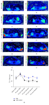
Results: The body weight growth rates in the SPIO-BMSC group were significantly higher than those in the PBS group (P < 0.05), and the Scr and BUN levels were also significantly lower than those in the PBS group (P < 0.05). HE staining showed that the degree of degeneration and vacuole-like changes in the renal tubular epithelial cells in the SPIO-BMSC group was significantly better than that observed in the PBS group. The TUNEL assay showed that the number of apoptotic renal tubular epithelial cells in the SPIO-BMSC group was significantly lower than that in the PBS group. The T2 value of the renal lesion was the highest on day 1 after cell transplantation, and it gradually decreased with time in both the PBS and SPIO-BMSC groups but was always the lowest in the SPIO-BMSC group.

Conclusion: SPIO-labeled BMSC transplantation can significantly promote the recovery of RIRI and noninvasive dynamic monitoring of engrafted cells and can also be performed simultaneously with MRI in vivo for the early prediction of therapeutic effects.


Figures
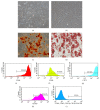
Figure 1
Figure 1
Passage 1 BMSCs on day 7 after isolation (a) and passage 3 BMSCs (b). Magnification, ×40 (a-b). Scale bar, 100 μm (a-b). Alizarin Red staining (c) and Oil Red O staining (d) of BMSCs after induced differentiation of osteogenesis and adipogenesis, respectively. Magnification, ×100 (c-d). Scale bar, 100 μm (c-d). The surface markers of the BMSCs were positive for CD44 (e), CD90 (f), CD73 (g), and CD105 (h) and negative for CD34, CD45, CD11b, CD19, and HLA-DR (i).
Figure 2
Figure 2
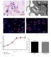
Prussian blue staining showing multiple blue particles (black arrow) in the cytoplasm of SPIO-labeled BMSCs (a). Magnification, ×400. Scale bar, 200 μm. TEM images showed many clusters of black particles (black arrow) in the cytoplasm of SPIO-labeled BMSCs (b). Scale bar, 200 nm. BrdU staining of unlabeled (c) and SPIO-labeled (d) BMSCs. Magnification, ×100 (c-d). Scale bar, 100 μm (c-d). Growth curves of unlabeled and SPIO-labeled BMSCs (e). Bar graph of BrdU-positive cells, with number of unlabeled and SPIO-labeled BMSCs (f).
Figure 3
Figure 3
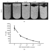
T2-weighted images of SPIO-labeled BMSCs labeled with various different cells numbers in vitro (0, 103, 105, 106, 2 × 10⁶) (a). Curve of T2 value for SPIO-labeled BMSCs changes with cells numbers (b).
Figure 4
Figure 4
Preparation of an RIRI model and BMSC transplantation. Before the renal artery was clamped (a). Forty-five minutes after the renal artery was clamped (b). 5 minutes (c) and 10 minutes (d) after the clamp was removed. HE staining of kidney tissue at 1 week after RIRI (e). Magnification, ×400. Scale bar, 200 μm. SPIO-labeled BMSCs were transplanted via the renal artery (f).
Figure 5
Figure 5
HE staining of kidney tissue on day 21 after cell transplantation in the SO group (a), PBS group (b), and SPIO-BMSC group (c). Magnification, ×100 (A-C). Scale bar, 100 μm (A–C). A bar graph of growth rate (d), Scr (e), and BUN (f) from each group on day 21 after cell transplantation.
Figure 6
Figure 6
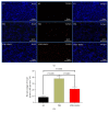
TUNEL staining of kidney tissue on day 21 after cell transplantation in each group (a). Magnification, ×100. Scale bar, 2 μm. A bar graph of the percentage of TUNEL-positive cells per mm2 from each group on day 21 after cell transplantation (b), 8.75±1.51% (SO group), 38.33±3.51% (PBS group), and 21.86±2.67% (SPIO-BMSC group).
Figure 7
Figure 7
T2WI of rat kidney in the PBS group before RIRI, on day 4 after RIRI, and on days 1, 7, and 21 after cells transplantation (a, c, e, g, i). T2WI of rat kidney in SPIO-BMSC group before RIRI, on day 4 after RIRI, and on days 1, 7, and 21 after cells transplantation (b, d, f, h, j). T2 value time curve of the renal lesion in the PBS and SPIO-BMSC groups (k).

Similar articles

Cited by

References

    1. Semedo P., Wang P. M., Andreucci T. H., et al. Mesenchymal stem cells ameliorate tissue damages triggered by renal ischemia and reperfusion injury. Transplantation Proceedings. 2007;39(2):421–423. doi: 10.1016/j.transproceed.2007.01.036. - DOI - PubMed
    1. Zhou L., Xu L., Shen J., et al. Preischemic administration of nonexpanded adipose stromal vascular fraction attenuates acute renal ischemia/reperfusion injury and fibrosis. Stem Cells Translational Medicine. 2016;5(9):1277–1288. doi: 10.5966/sctm.2015-0223. - DOI - PMC - PubMed
    1. Kale S., Karihaloo A., Clark P. R., Kashgarian M., Krause D. S., Cantley L. G. Bone marrow stem cells contribute to repair of the ischemically injured renal tubule. The Journal of Clinical Investigation. 2003;112(1):42–49. doi: 10.1172/JCI200317856. - DOI - PMC - PubMed
    1. Morigi M., Introna M., Imberti B. Human bone marrow mesenchymal stem cells accelerate recovery of acute renal injury and prolong survival in mice. Stem Cells. 2008;26(8):2075–2082. doi: 10.1634/stemcells.2007-0795. - DOI - PubMed
    1. Council N. (85-23(rev.)).Guide for the Care and Use of Laboratory Animals. (8th) 2011;327 doi: 10.17226/12910. - DOI

MeSH terms

LinkOut - more resources