

Radiological findings of Posterior Reversible Encephalopathy Syndrome in transplanted children previous affected by hemoglobinopathy: A neuroimaging retrospective analysis
- PMID: 31016209
- PMCID: PMC6468159
- DOI: 10.1016/j.ejro.2019.03.001
Radiological findings of Posterior Reversible Encephalopathy Syndrome in transplanted children previous affected by hemoglobinopathy: A neuroimaging retrospective analysis
Abstract
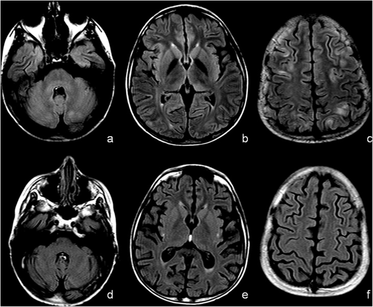
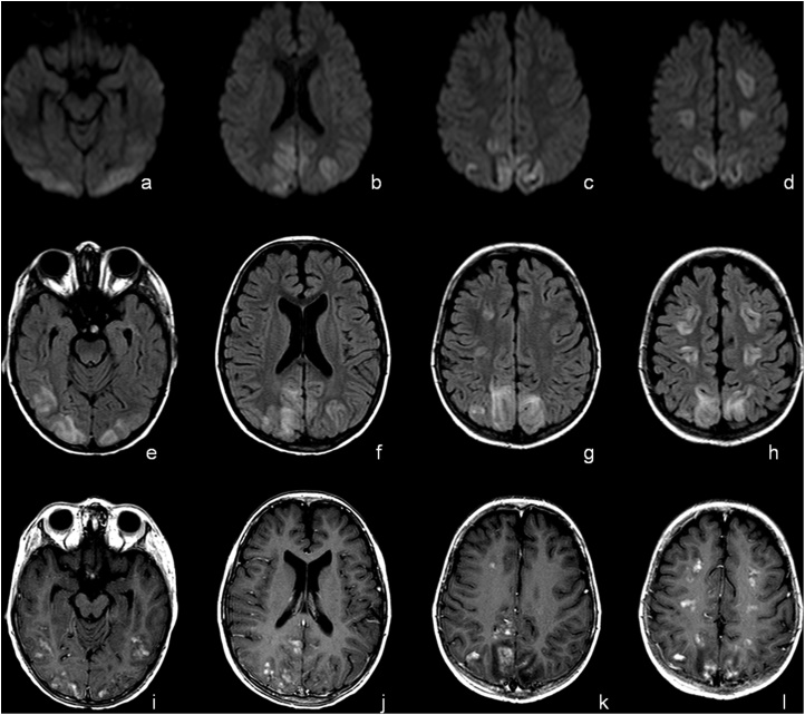
To evaluate, by Magnetic Resonance Imaging, if there is a typical pattern or severity of PRES in transplanted children for hemoglobinopathy. Secondary point was to investigate the pattern and severity of PRES in children with thalassemia-THAL and sickle-cell disease-SCD after autologous hematopoietic stem cell transplantation (aHSCT). Finally, we evaluate the presence of atypical PRES presentation and the involved area of central nervous system. Two neuroradiologists analyzed retrospectively MRI of 21 transplanted children for THAL or SCD treated with CI, with neurological symptoms and signs of PRES. The Bartynski and Boardman classification has been used for PRES pattern while McKinney scale for PRES severity. Fisher Exact Probability test or Chi-square test were used to compare the categorical data. In the 21 transplanted children the PRES severity was typically mild (85.7%) without preferring radiological pattern at MRI. The analysis didn't show significant association between PRES pattern or PRES severity and previous hemoglobinopathy (THAL or SCD). No atypical PRES presentation has been found. PRES severity in transplanted children for hemoglobinopathy is typically mild. Notwithstanding children affected by SCD have a damage on the capillary endothelium, after aHSCT our data didn't show a different PRES severity and pattern than THAL children.
Keywords: Hemoglobinopathy; MRI; PRES; Sickle-cell disease; Thalassemia; aHSCT.
Figures


Similar articles
-
Posterior Reversible Encephalopathy Syndrome after Hematopoietic Cell Transplantation in Children with Hemoglobinopathies.Biol Blood Marrow Transplant. 2017 Sep;23(9):1531-1540. doi: 10.1016/j.bbmt.2017.05.033. Epub 2017 Jul 3. Biol Blood Marrow Transplant. 2017. PMID: 28602890
-
Analysis of Risk Factors Associated With Poor Outcome in Posterior Reversible Encephalopathy Syndrome After Treatment in Children: Systematic Review and Meta-Analysis.Front Neurol. 2020 Aug 26;11:938. doi: 10.3389/fneur.2020.00938. eCollection 2020. Front Neurol. 2020. PMID: 32982945 Free PMC article.
-
Neurotoxicity including posterior reversible encephalopathy syndrome after initiation of calcineurin inhibitors in transplanted methylmalonic acidemia patients: Two case reports and review of the literature.JIMD Rep. 2020 Jan 22;51(1):89-104. doi: 10.1002/jmd2.12088. eCollection 2020 Jan. JIMD Rep. 2020. PMID: 32071844 Free PMC article.
-
Mixed chimerism following in utero hematopoietic stem cell transplantation in murine models of hemoglobinopathy.Exp Hematol. 2003 Feb;31(2):176-84. doi: 10.1016/s0301-472x(02)01024-x. Exp Hematol. 2003. PMID: 12591283
-
Neurological PRESentations in Sickle Cell Patients Are Not Always Stroke: A Review of Posterior Reversible Encephalopathy Syndrome in Sickle Cell Disease.Pediatr Blood Cancer. 2016 Jun;63(6):983-9. doi: 10.1002/pbc.25932. Epub 2016 Feb 12. Pediatr Blood Cancer. 2016. PMID: 26871763 Review.
Cited by
-
Bithalamic infarction in a tentorial dural artero-venous fistula and thalamic dementia: a case report and systematic review.Neurol Sci. 2023 Jul;44(7):2291-2304. doi: 10.1007/s10072-023-06716-w. Epub 2023 Mar 18. Neurol Sci. 2023. PMID: 36932275
-
Posterior reversible encephalopathy syndrome and Wernicke encephalopathy in patient with acute graft-versus-host disease.Radiol Case Rep. 2019 May 31;14(8):971-976. doi: 10.1016/j.radcr.2019.05.024. eCollection 2019 Aug. Radiol Case Rep. 2019. PMID: 31193981 Free PMC article.
-
Paediatric posterior reversible encephalopathy syndrome: is there an association of blood pressure with imaging severity and atypical magnetic resonance characteristics?Pediatr Radiol. 2022 Dec;52(13):2610-2619. doi: 10.1007/s00247-022-05400-z. Epub 2022 Jun 20. Pediatr Radiol. 2022. PMID: 35723697 Review.
-
Metagenomic Next Generation Sequencing in the Detection of Pathogens in Cerebrospinal Fluid of Patients After Alternative Donor Transplantation: A Feasibility Analysis.Front Cell Infect Microbiol. 2021 Sep 14;11:720132. doi: 10.3389/fcimb.2021.720132. eCollection 2021. Front Cell Infect Microbiol. 2021. PMID: 34595132 Free PMC article.
-
Qualitative and quantitative analysis of 3D T1 Silent imaging.Radiol Med. 2021 Sep;126(9):1207-1215. doi: 10.1007/s11547-021-01380-6. Epub 2021 Jun 15. Radiol Med. 2021. PMID: 34131844
References
-
- Lucarelli G., Gaziev J. Advances in the allogeneic transplantation for thalassemia. Blood Rev. 2008;22:53–63. - PubMed
-
- Gaziev J. Optimal outcomes in young class 3 patients with thalassemia undergoing HLA-identical sibling bone marrow transplantation. Transplantation. 2016;100:925–932. - PubMed
-
- McKinney A.M. Posterior reversible encephalopathy syndrome: incidence of atypical regions of involvement and imaging findings. Am J Roentgenol. 2007;189:904–912. - PubMed
LinkOut - more resources
Full Text Sources
