Case Reports
. 2019 Apr 16:5:12.
doi: 10.1038/s41523-019-0107-9. eCollection 2019.

Initial experience of dedicated breast PET imaging of ER+ breast cancers using [F-18]fluoroestradiol

Affiliations
Case Reports

Initial experience of dedicated breast PET imaging of ER+ breast cancers using [F-18]fluoroestradiol

Ella F Jones et al. NPJ Breast Cancer. .

Abstract

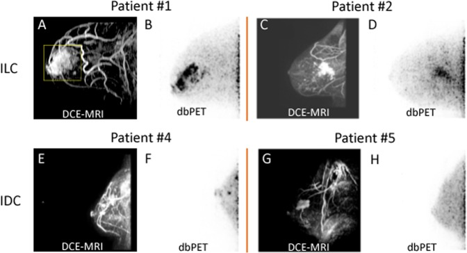
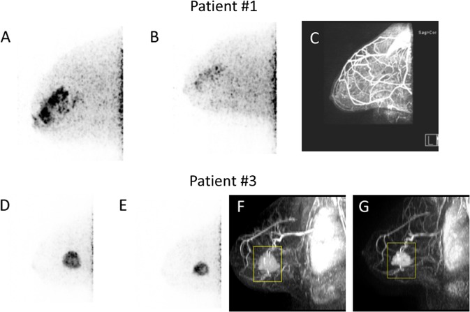
Dedicated breast positron emission tomography (dbPET) is an emerging technology with high sensitivity and spatial resolution that enables detection of sub-centimeter lesions and depiction of intratumoral heterogeneity. In this study, we report our initial experience with dbPET using [F-18]fluoroestradiol (FES) in assessing ER+ primary breast cancers. Six patients with >90% ER+ and HER2- breast cancers were imaged with dbPET and breast MRI. Two patients had ILC, three had IDC, and one had an unknown primary tumor. One ILC patient was treated with letrozole, and another patient with IDC was treated with neoadjuvant chemotherapy without endocrine treatment. In this small cohort, we observed FES uptake in ER+ primary breast tumors with specificity to ER demonstrated in a case with tamoxifen blockade. FES uptake in ILC had a diffused pattern compared to the distinct circumscribed pattern in IDC. In evaluating treatment response, the reduction of SUVmax was observed with residual disease in an ILC patient treated with letrozole, and an IDC patient treated with chemotherapy. Future study is critical to understand the change in FES SUVmax after endocrine therapy and to consider other tracer uptake metrics with SUVmax to describe ER-rich breast cancer. Limitations include variations of FES uptake in different ER+ breast cancer diseases and exclusion of posterior tissues and axillary regions. However, FES-dbPET has a high potential for clinical utility, especially in measuring response to neoadjuvant endocrine treatment. Further development to improve the field of view and studies with a larger cohort of ER+ breast cancer patients are warranted.


Conflict of interest statement

The authors declare no competing interests.

Figures

Fig. 1
Fig. 1
Examples of FES uptake in >90% ER+/HER2− invasive lobular carcinomas (ILC, top panel) and invasive ductal carcinomas (IDC, bottom panel). Top—a Patient #1—a 61-year-old female patient with grade 2 ILC in her right breast. MRI showed contrast enhancement spanning 6.7 cm. b FES-dbPET showed an SUVmax at 15.83 and total uptake volume at 15.72 cm3. c Patient #2—a 64-year-old female patient with grade 2 ILC in her right breast. MRI showed an irregularly shaped mass with spiculated margins associating with non-mass enhancement of 5.3 cm. d The corresponding FES-dbPET showed a SUVmax at 22.42 and total uptake volume at 35.63 cm3. Bottom—e Patient #4—a 63-year-old female patient with grade 2 IDC in her left breast. MRI showed a 0.9 cm confirmed IDC and a 1.3 cm irregular mass with posterior depth. f FES-dbPET showed a SUVmax at 7.44 corresponding to the 0.9 cm anterior mass but missed the posterior mass that was close to the chest wall. g Patient #5—a 33-year-old female patient with grade 2 IDC in her right breast. MRI showed an enhancing mass with spiculated margin spanning 3.4 cm. h The patient was treated with tomoxifen for fertility preservation and stopped 4 days prior to FES-dbPET imaging. The corresponding FES-dbPET showed no FES uptake that was consistent with ER blockade
Fig. 2
Fig. 2
Examples of FES-dbPET for assessing treatment response. Top patient #1 with ILC—a FES-dbPET imaging at baseline with a SUVmax at 15.83 and total uptake volume at 15.72 cm3. b FES-dbPET of the same patient after 2 months of treatment with letrozole showing a SUVmax at 6.11 and total uptake volume at 0.37 cm3. c DCE-MRI after 3 months of treatment with letrozole, confirming the favorable response with no residual disease but with significant background enhancement. Bottom patient #3 with IDC—d A 49-year-old female patient with grade 2 IDC in her left breast. FES-dbPET imaging at baseline showed a SUVmax at 13.02 and total uptake volume at 12.60 cm3. e FES-dbPET of the same patient after 3 weeks of treatment with chemotherapy without endocrine therapy had a SUVmax at 16.26 but a reduced total uptake volume at 6.97 cm3 (45% reduction). f, g DCE-MRI at baseline and after 3 weeks of treatment, confirming the tumor size reduction by 40%

Similar articles

Cited by

References

    1. Lippman ME, et al. The relation between estrogen receptors and response rate to cytotoxic chemotherapy in metastatic breast cancer. N. Engl. J. Med. 1978;298:1223–1228. doi: 10.1056/NEJM197806012982203. - DOI - PubMed
    1. McGuire WL, Horwitz KB, Pearson OH, Segaloff A. Current status of estrogen and progesterone receptors in breast cancer. Cancer. 1977;39:2934–2947. doi: 10.1002/1097-0142(197706)39:6<2934::AID-CNCR2820390680>3.0.CO;2-P. - DOI - PubMed
    1. Sparano JA, et al. Prospective validation of a 21-gene expression assay in breast cancer. N. Eng. J. Med. 2015;373:2005–2014. doi: 10.1056/NEJMoa1510764. - DOI - PMC - PubMed
    1. Stearns V. TAILORing adjuvant systemic therapy for breast cancer. N. Engl. J. Med. 2018;379:191–192. doi: 10.1056/NEJMe1806329. - DOI - PubMed
    1. Exner, R. et al. The multigene signature MammaPrint impacts on multidisciplinary team decisions in ER+, HER2- early breast cancer. Br. J. Cancer111, 837 (2014). - PMC - PubMed

Publication types