. 2019 Jun;23(6):4097-4110.
doi: 10.1111/jcmm.14297. Epub 2019 Apr 23.

TSP-1 is downregulated and inversely correlates with miR-449c expression in Cushing's disease

Affiliations

TSP-1 is downregulated and inversely correlates with miR-449c expression in Cushing's disease

Jie Ren et al. J Cell Mol Med. 2019 Jun.

Abstract

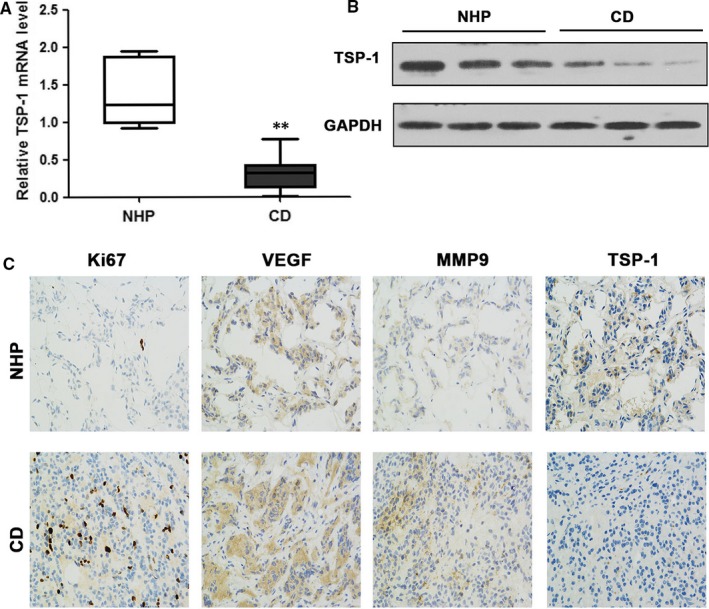
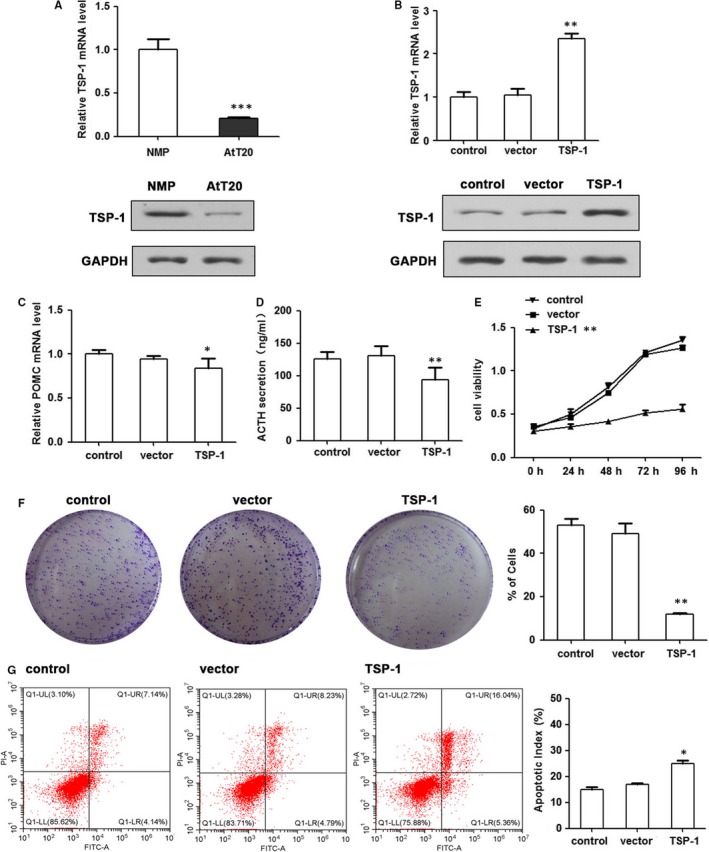
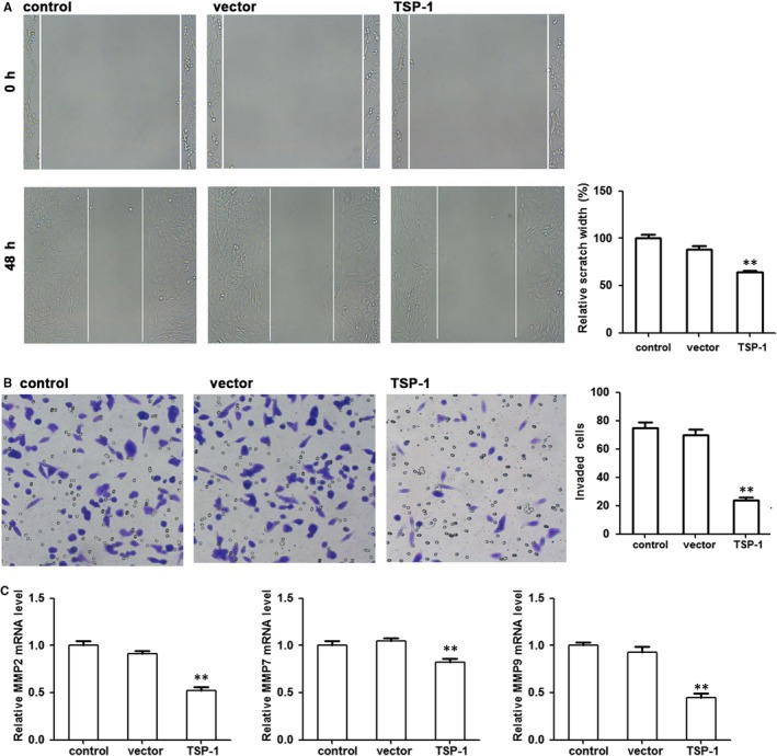
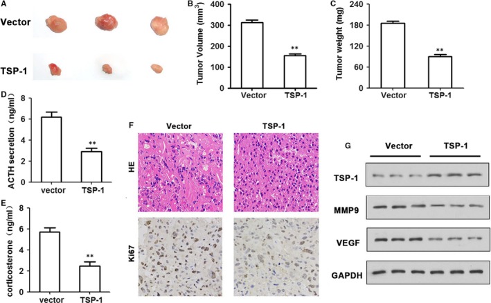
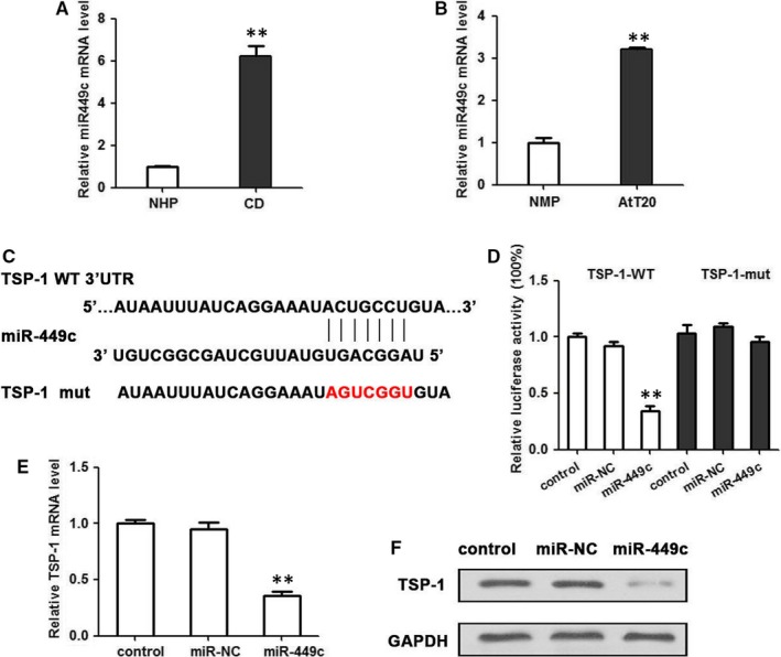
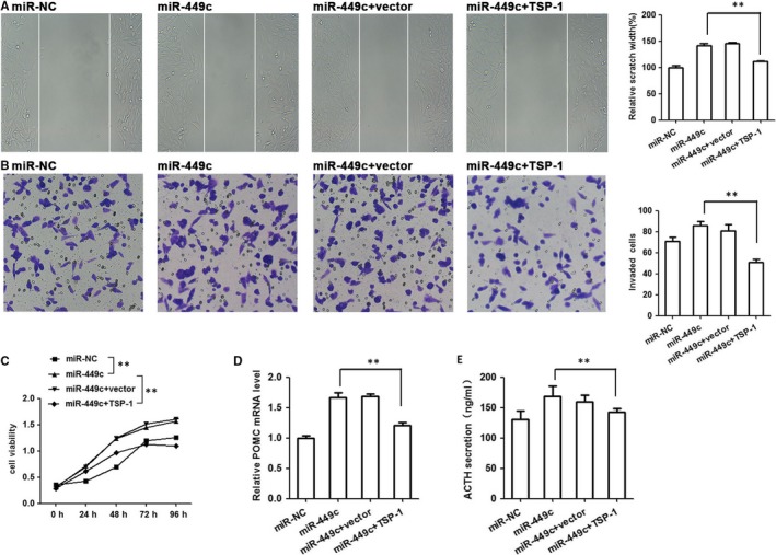
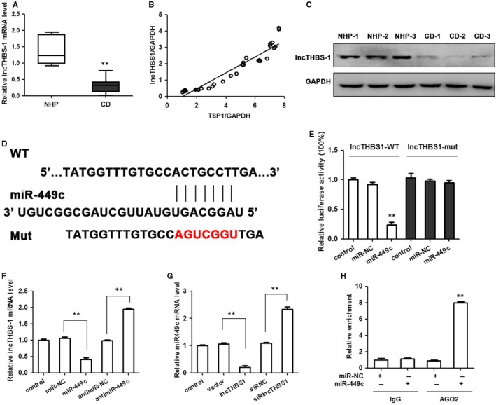
The pathogenesis of Cushing's disease, which is caused by pituitary corticotroph adenoma, remains to be studied. Secreted angioinhibitory factor thrombospondin-1 (TSP-1) is an adhesive glycoprotein that mediates cell-to-cell and cell-to-matrix interactions and is associated with platelet aggregation, angiogenesis and tumorigenesis. We have found that the expression of TSP-1 is significantly lower in human pituitary corticotroph tumours compared with normal adenohypophysis. This study aims to elucidate the role of TSP-1 in regulating the tumour function of pituitary adenomas. Forced overexpression of TSP-1 in a murine AtT20 pituitary corticotroph tumour cell line decreased corticotroph precursor hormone proopiomelanocortin (POMC) transcription and adrenocorticotropic hormone (ACTH) secretion. Functional studies showed that TSP-1 overexpression in pituitary adenoma cells suppressed proliferation, migration and invasion. We have demonstrated that TSP-1 is a direct target of miR-449c. Further study showed that miR-449c activity enhanced tumorigenesis by directly inhibiting TSP-1 expression. Low expression of lncTHBS1, along with low expression of TSP-1, was associated with the high expression of miR-449c in Cushing's disease patients. Furthermore, RNA-immunoprecipitation associates miR-449c with lncTHBS1 suggesting that lncTHBS1 might be a negative regulator of miR-449c. Taken together, this study has demonstrated that lncTHBS1 might function as competing endogenous RNA for miR-449c, which could suppress the development of Cushing's disease.

Keywords: ACTH-secreting pituitary adenomas; Cushing's disease; lncTHBS1; miR-449c; thrombospondin-1.


Conflict of interest statement

The authors confirm that there are no conflicts of interest.

Figures

Figure 1
Figure 1
Relative TSP‐1 expression in pituitary corticotrophs. A, The expression of TSP‐1 mRNA in Cushing's disease (CD, n = 33) and normal human pituitary (NHP, n = 7) tissue. B, Western blotting of TSP‐1 protein levels in normal pituitary and CD tissue samples. C, Immunohistochemical staining for Ki67, VEGF, MMP9 and TSP‐1 in representative normal pituitary and corticotroph adenoma (magnification, ×200). Representations of at least three biological replicates are presented (mean ± SEM; **P < 0.01)
Figure 2
Figure 2
TSP‐1 decreases the proliferation and clonogenic ability of AtT20 cells in vitro. Overexpression of TSP‐1 levels through the transfection of pcDNA‐TSP‐1 in AtT20. A, The relative expression of TSP‐1 was measured by qRT‐PCR and WB. B, qRT‐PCR and WB assay confirmed increased expression of TSP‐1 in stably transfected AtT20 cells. C, TSP‐1 effects on POMC expression was detected by qRT‐PCR (n = 3; *P < 0.05, **P < 0.01, ***P < 0.001 vs control). D, TSP‐1 effects on adrenocorticotropic hormone (ACTH) secretion was detected by ELISA assay. E, MTT assay showed that overexpression TSP‐1 decreased cell growth rates. F, Colony formation was assessed under a microscope. (n = 3, *P < 0.05, **P < 0.01). G, Cell apoptosis was determined by flow cytometry. Mean ± SEM; *P < 0.05, **P < 0.01, ***P < 0.001
Figure 3
Figure 3
TSP‐1 inhibits AtT20 cell migration. A, Representative images of the wound healing assay in the TSP‐1‐transfected or vector‐transfected AtT20 cells at 48 h. B, Transwell invasion assay was employed to assess the invasive potential of overexpressing TSP‐1 in AtT20 cells. C, Relative expression of MMP2, MMP7 and MMP9 by qRTPCR. **P < 0.01
Figure 4
Figure 4
TSP‐1 inhibits tumour growth in vivo. A, TSP‐1‐overexpressed AtT20 cells were injected subcutaneously in the nude mice. Mice were killed 21 d later for evaluation. Gross morphology of tumour is shown. B, TSP‐1 upregulation decreased the volume of tumours compared with the control group (n = 6 per group). (D‐E) ACTH (D) and corticosterone (E) levels derived from mice harbouring stable TSP‐1 overexpressing cells compared with controls by ELISA assay. F, Hematoxylin‐eosin staining of samples and immunoreactivity to Ki67 as observed by fluorescence microscopy (magnification ×400). G, The protein expression of TSP‐1, VEGF and MMP9 was measured by Western blotting. GAPDH was used as an internal control. Representations of at least three biological replicates are presented (mean ± SEM; **P < 0.01)
Figure 5
Figure 5
miR‐449c is expressed in pituitary corticotroph tumours and targets the 3′UTR of TSP‐1. A, The expression of miR‐449c mRNA in Cushing's disease (n = 33) and normal pituitary tissue (n = 7) were determined by qRT‐PCR. **P < 0.01. B, The expression of miR‐449c mRNA in AtT20 cells and normal mouse pituitary, tested by qRT‐PCR. C, The predicted target sequence of miR‐449c in the 3′UTR of TSP‐1 and mutant containing three altered nucleotides in the seed sequence of miR‐449c. D, Luciferase assay of pGL3‐TSP‐1WT or pGL3‐TSP‐1 mutant in the presence of miR‐449c mimic in the AtT20 cell line. Luciferase activity was detected 48 h after transfection and normalized to Renilla. (E) qRT‐PCR and (F) Western blotting analysis of miR‐449c effects on mRNA level of TSP‐1 in AtT20 cells. **P < 0.01
Figure 6
Figure 6
TSP‐1 reversed miR‐449c expression to affect the function of AtT20 cells in vitro. AtT20 cells were transfected with mimics control or miR‐449c and miR‐449c+TSP1. A, Invasion of AtT20 cells in different groups by wound healing assay. B, Transwell invasion assay, (C) MTT assay. (D) qRT‐PCR analysis of miR‐449c and TSP‐1 effects on POMC expression, and (E) miR‐449c and TSP‐1 effects on ACTH secretion. **P < 0.01
Figure 7
Figure 7
Positive correlation between TSP‐1 and lncTHBS1 expression. lncTHBS1 targets miR‐449c by directly binding to a miRNA response element. A, The expression of lncTHBS1 mRNA in CD (n = 33) and normal pituitary tissue (n = 7). B, Correlation analysis of the relationship between lncTHBS1 expression and TSP‐1 level. C, Northern blot assays were used to detect lncTHBS1 expression in CD. D, Schematic representation of the predicted binding sites for miR‐449c, and the site mutagenesis design for the reporter assay. E, The relative luciferase activities were inhibited in the HEK‐293T cells transfected with the reporter vector lncTHBS1‐WT, but not with the reporter vector lncTHBS1‐Mut. F, miR‐449c expression decreased lncTHBS1 expression, and inhibition of miR‐449c enhanced lncTHBS1 expression in the HEK‐293T cells. G, lncTHBS1 expression decreased miR‐449c expression whereas inhibition of lncTHBS1 increased miR‐449c expression in HEK‐293T cells. H, miR‐449c was identified in the lncTHBS1 complex. miR‐NC and miR‐449c cell lysates were used for RNA‐IP with anti‐Ago2 antibody. Cells transfected with miR‐449c mimics or miR‐NC, followed by qRT‐PCR to detect lncTHBS1. **P < 0.01

Similar articles

Cited by

References

    1. Colao A, Boscaro M, Ferone D, Casanueva FF. Managing Cushing's disease: the state of the art. Endocrine. 2014;47:9‐20. - PubMed
    1. Biller BM, Grossman AB, Stewart PM, et al. Treatment of adrenocorticotropin‐dependent Cushing's syndrome: a consensus statement. J Clin Endocrinol Metab. 2008;93:2454‐2462. - PMC - PubMed
    1. Pivonello R, De Martino MC, De Leo M, et al. Cushing's syndrome. Endocrinol Metab Clin North Am. 2008;37:135-149. - PubMed
    1. Praw SS, Heaney AP. Medical treatment of Cushing's disease: overview and recent findings. Int J Gen Med. 2009;2:209‐217. - PMC - PubMed
    1. Martin R, Cetin Y, Fehm HL, Fahlbusch R, Voigt KH. Multiple cellular forms of corticotrophs in surgically removed pituitary adenomas and periadenomatous tissue in Cushing's disease. Am J Pathol. 1982;106:332‐341. - PMC - PubMed

Publication types