

Role of the thioredoxin interacting protein in diabetic nephropathy and the mechanism of regulating NOD‑like receptor protein 3 inflammatory corpuscle
- PMID: 31017263
- PMCID: PMC6488169
- DOI: 10.3892/ijmm.2019.4163
Role of the thioredoxin interacting protein in diabetic nephropathy and the mechanism of regulating NOD‑like receptor protein 3 inflammatory corpuscle
Abstract
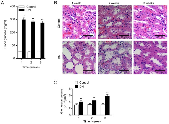
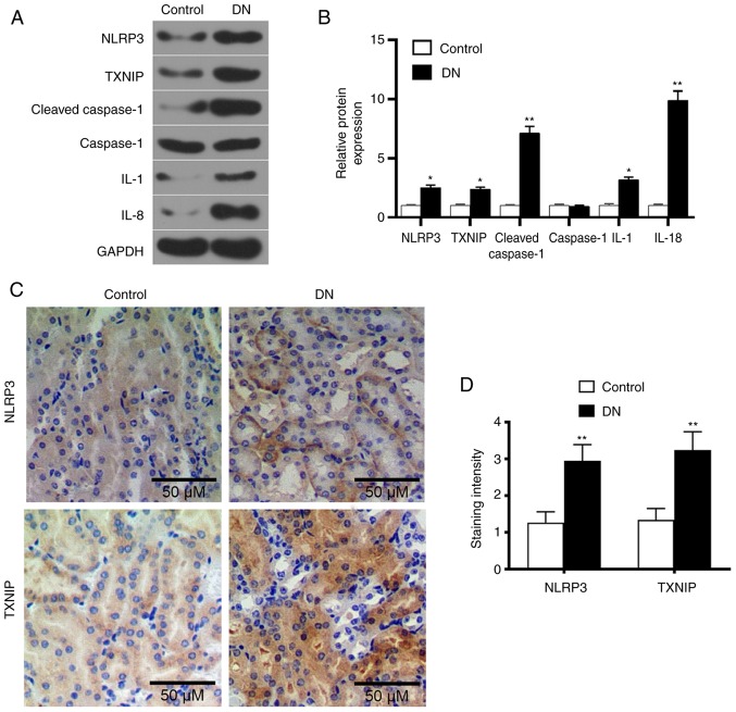
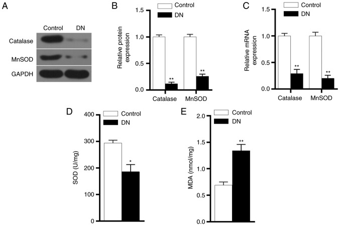
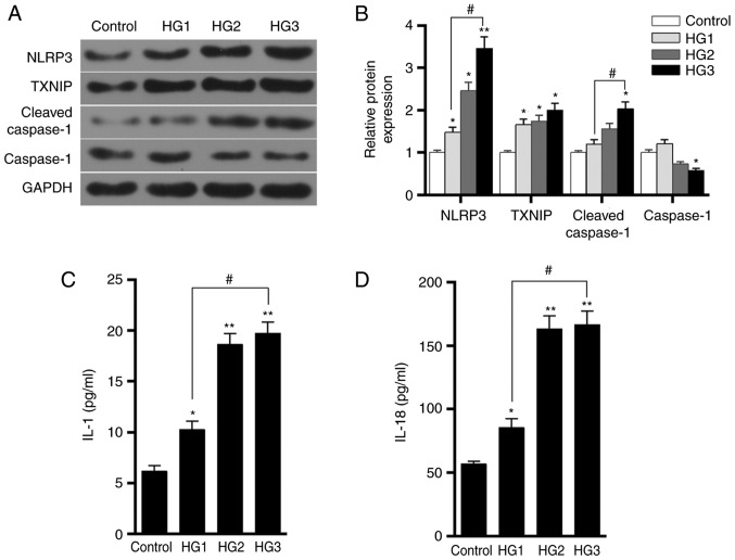
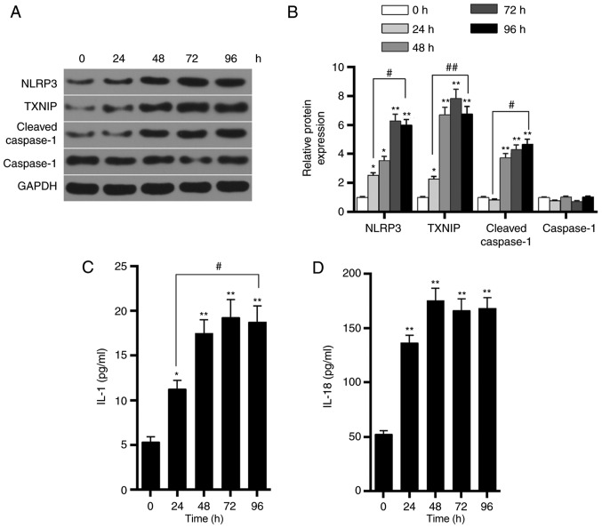
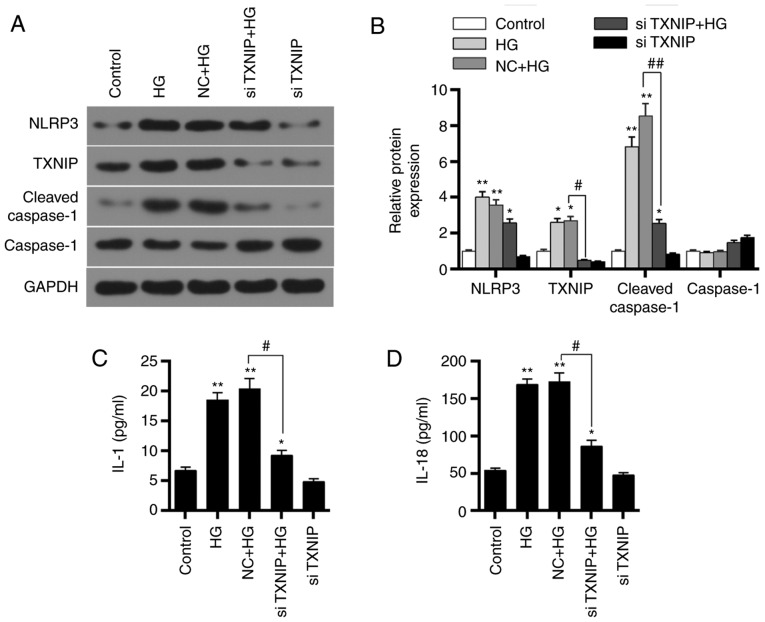
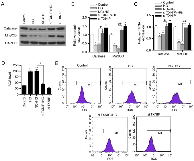
Inflammatory response serves an important role in diabetic nephropathy (DN); however, the mechanism of inflammatory response results in renal damage is not yet clear. The aim of the present study was to investigate the role of the thioredoxin interacting protein (TXNIP)/NOD‑like receptor protein 3 (NLRP3) axis‑mediated activation of NLRP3 inflammasome in DN. A diabetic rat model was induced by streptozotocin injection. Hematoxylin and eosin (H&E) staining and streptavidin‑peroxidase staining were then used to examine the kidney tissue morphology, and TXNIP and NLRP3 protein expression levels, respectively. Furthermore, RNA interference technology was applied to silence the TXNIP gene. TXNIP and NLRP3 inflammasome activation‑associated proteins and mRNAs were detected by western blot analysis and RT‑qPCR, respectively. Enzyme‑linked immunosorbent assay was also used to examine interleukin (IL)‑1 and IL‑18 expression, while flow cytometry was performed to detect reactive oxygen species production. The data revealed that TXNIP and NLRP3 were overexpressed in kidney tissue of DN rats, and the level of antioxidant genes was downregulated. It was also observed that glucose promoted TXNIP expression and activation of NLRP3 inflammasome in a time‑dependent and dose‑dependent manner, therefore promoting inflammatory responses. Silencing of TXNIP gene resulted in the downregulation of NLRP3 inflammasome activation, and inhibited the expression levels of IL‑1 and IL‑18 in a high‑glucose environment. Furthermore, low expression of TXNIP promoted the levels of antioxidant genes and reduced the ROS levels. Taken together, the high‑glucose environment was able to upregulated the level of inflammatory factors by promoting the expression of TXNIP and the activation of NLRP3 inflammasome, consequently participating in DN.
Figures
Similar articles
-
Activation of the TXNIP/NLRP3 inflammasome pathway contributes to inflammation in diabetic retinopathy: a novel inhibitory effect of minocycline.Inflamm Res. 2017 Feb;66(2):157-166. doi: 10.1007/s00011-016-1002-6. Epub 2016 Oct 26. Inflamm Res. 2017. PMID: 27785530
-
NLRP3 deficiency ameliorates renal inflammation and fibrosis in diabetic mice.Mol Cell Endocrinol. 2018 Dec 15;478:115-125. doi: 10.1016/j.mce.2018.08.002. Epub 2018 Aug 8. Mol Cell Endocrinol. 2018. PMID: 30098377
-
Thioredoxin-Interacting Protein Mediates NLRP3 Inflammasome Activation Involved in the Susceptibility to Ischemic Acute Kidney Injury in Diabetes.Oxid Med Cell Longev. 2016;2016:2386068. doi: 10.1155/2016/2386068. Epub 2016 Oct 27. Oxid Med Cell Longev. 2016. PMID: 27867451 Free PMC article.
-
Roles of the NLRP3 inflammasome in the pathogenesis of diabetic nephropathy.Pharmacol Res. 2016 Dec;114:251-264. doi: 10.1016/j.phrs.2016.11.004. Epub 2016 Nov 5. Pharmacol Res. 2016. PMID: 27826011 Review.
-
Effects of antidiabetic drugs on NLRP3 inflammasome activity, with a focus on diabetic kidneys.Drug Discov Today. 2019 Jan;24(1):256-262. doi: 10.1016/j.drudis.2018.08.005. Epub 2018 Aug 4. Drug Discov Today. 2019. PMID: 30086405 Review.
Cited by
-
Comprehensive Diagnostics of Diabetic Nephropathy by Transcriptome RNA Sequencing.Diabetes Metab Syndr Obes. 2022 Oct 10;15:3069-3080. doi: 10.2147/DMSO.S371026. eCollection 2022. Diabetes Metab Syndr Obes. 2022. PMID: 36237968 Free PMC article.
-
Epigallocatechin-3-Gallate Ameliorates Diabetic Kidney Disease by Inhibiting the TXNIP/NLRP3/IL-1β Signaling Pathway.Food Sci Nutr. 2024 Nov 26;12(12):10800-10815. doi: 10.1002/fsn3.4617. eCollection 2024 Dec. Food Sci Nutr. 2024. PMID: 39723074 Free PMC article.
-
Canagliflozin Attenuates Podocyte Inflammatory Injury through Suppressing the TXNIP/NLRP3 Signaling Pathway in Diabetic Kidney Disease Mice.Inflammation. 2025 Mar 11. doi: 10.1007/s10753-025-02258-9. Online ahead of print. Inflammation. 2025. PMID: 40067577
-
New Insights into the Mechanisms of Pyroptosis and Implications for Diabetic Kidney Disease.Int J Mol Sci. 2020 Sep 25;21(19):7057. doi: 10.3390/ijms21197057. Int J Mol Sci. 2020. PMID: 32992874 Free PMC article. Review.
-
Reactive Oxygen Species: Drivers of Physiological and Pathological Processes.J Inflamm Res. 2020 Dec 2;13:1057-1073. doi: 10.2147/JIR.S275595. eCollection 2020. J Inflamm Res. 2020. PMID: 33293849 Free PMC article. Review.
References
MeSH terms
Substances
LinkOut - more resources
Full Text Sources
Medical
Miscellaneous
