. 2020 Feb;177(4):793-809.
doi: 10.1111/bph.14691. Epub 2019 Apr 24.

Hydrogen sulfide inhibits calcification of heart valves; implications for calcific aortic valve disease

Affiliations

Hydrogen sulfide inhibits calcification of heart valves; implications for calcific aortic valve disease

Katalin Éva Sikura et al. Br J Pharmacol. 2020 Feb.

Abstract

Background and purpose: Calcification of heart valves is a frequent pathological finding in chronic kidney disease and in elderly patients. Hydrogen sulfide (H2 S) may exert anti-calcific actions. Here we investigated H2 S as an inhibitor of valvular calcification and to identify its targets in the pathogenesis.

Experimental approach: Effects of H2 S on osteoblastic transdifferentiation of valvular interstitial cells (VIC) isolated from samples of human aortic valves were studied using immunohistochemistry and western blots. We also assessed H2S on valvular calcification in apolipoprotein E-deficient (ApoE-/- ) mice.

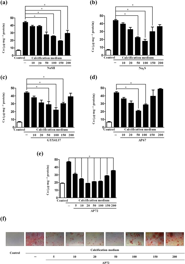
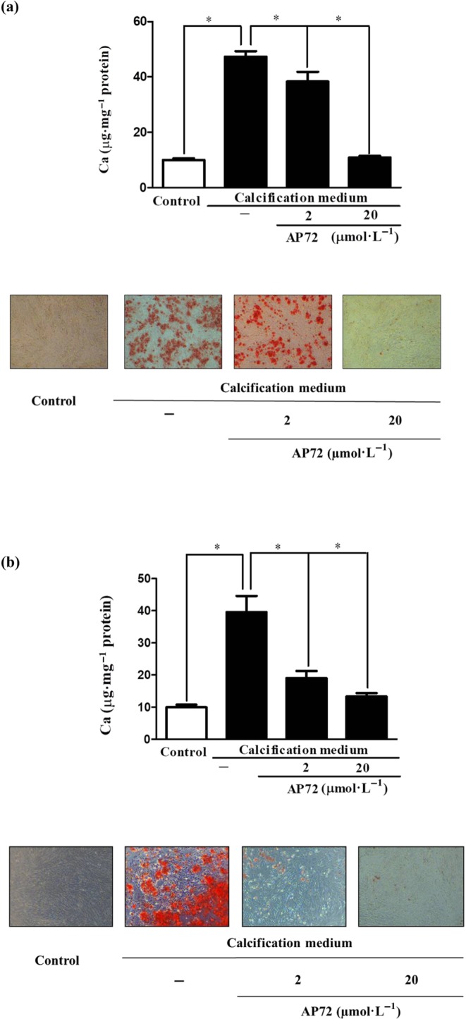
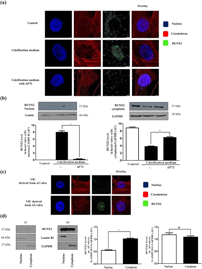
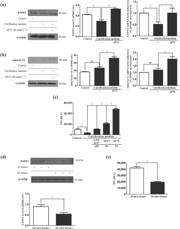
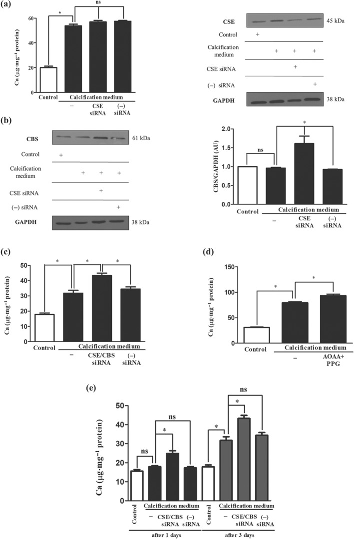
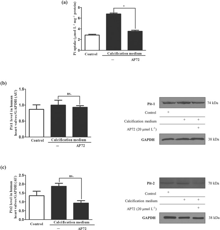
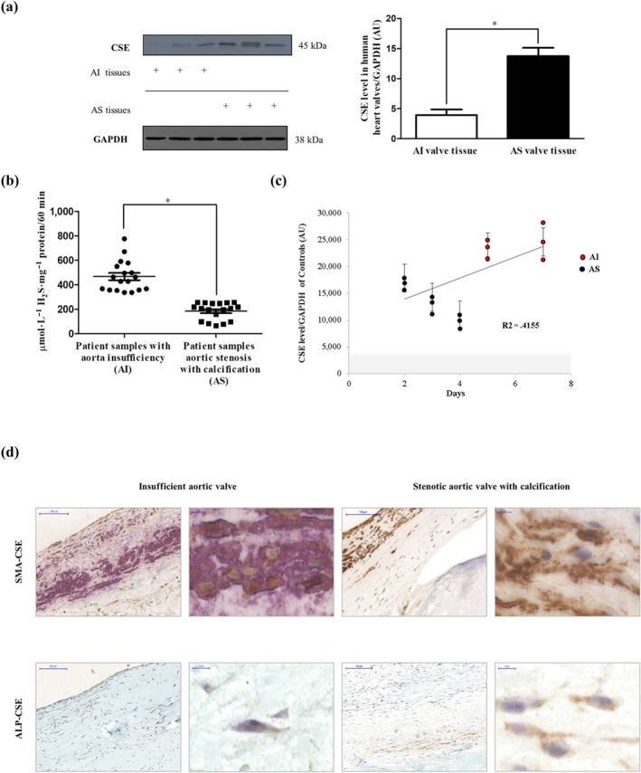
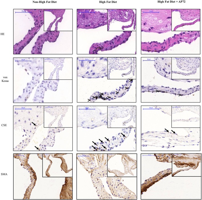
Key results: In human VIC, H2 S from donor compounds (NaSH, Na2 S, GYY4137, AP67, and AP72) inhibited mineralization/osteoblastic transdifferentiation, dose-dependently in response to phosphate. Accumulation of calcium in the extracellular matrix and expression of osteocalcin and alkaline phosphatase was also inhibited. RUNX2 was not translocated to the nucleus and phosphate uptake was decreased. Pyrophosphate generation was increased via up-regulating ENPP2 and ANK1. Lowering endogenous production of H2 S by concomitant silencing of cystathionine γ-lyase (CSE) and cystathionine β-synthase (CBS) favoured VIC calcification. analysis of human specimens revealed higher Expression of CSE in aorta stenosis valves with calcification (AS) was higher than in valves of aortic insufficiency (AI). In contrast, tissue H2 S generation was lower in AS valves compared to AI valves. Valvular calcification in ApoE-/- mice on a high-fat diet was inhibited by H2 S.

Conclusions and implications: The endogenous CSE-CBS/H2 S system exerts anti-calcification effects in heart valves providing a novel therapeutic approach to prevent hardening of valves.

Linked articles: This article is part of a themed section on Hydrogen Sulfide in Biology & Medicine. To view the other articles in this section visit http://onlinelibrary.wiley.com/doi/10.1111/bph.v177.4/issuetoc.


Conflict of interest statement

The authors declare no conflicts of interest. M.W. has patent applications for the therapeutic use of slow‐release hydrogen sulfide donor molecules.

Figures

Figure 1
Figure 1
H2S donors dose‐dependently inhibit the calcification of VIC. Calcium contents of VIC in growth medium or calcification medium are shown, after 5 days treatment with (a) NaSH, (b) Na2S, (c) GYY4137, (d) AP67 (10–200 μmol·L−1), or with (e) AP72 (5–200 μmol·L−1). (f) Alizarin Red S staining of VIC. Data shown are means ± SEM of five independent experiments. *P < .05, significantly different as indicated
Figure 2
Figure 2
Phenol red impairs anti‐calcification effect of H2S. Cultured VIC in (a) phenol red‐containing DMEM (Sigma) or (b) phenol red‐free media were supplemented with AP72 (2 μmol·L−1; 20 μmol·L−1) for 5 days, and calcium content of the cells was measured and normalized to protein content of the cells. Alizarin Red S staining represents the microscopic image of calcium deposition of extracellular matrix. Data shown are means ± SEM of five independent experiments. *P < .05, significantly different as indicated
Figure 3
Figure 3
AP72 prevents nuclear translocation of RUNX2. VIC grown on coverslips were exposed to growth medium or calcification medium alone or supplemented with AP72 (20 μmol·L−1) for 2 days. (a) Cells were stained for DNA (Hoechst 33258, blue), RUNX2 (green, Alexa Flour 488), and F‐actin (cytoskeleton, red, iFluor 647). Images are obtained employing an immunofluorescence‐confocal STED nanoscope. (b) RUNX2 expression in cytoplasmic and nuclear fraction of VIC. The band intensities are normalized for Lamin B1 in case of nuclear extracts and for GAPDH in case of cytoplasm extracts. (c) RUNX2 localization in AI‐ and AS‐derived VIC samples. Images are obtained employing an immunofluorescence‐confocal STED nanoscope. (d) Protein levels of RUNX2 in AI and AS tissue lysates are shown. Representative staining and protein analysis are shown from five independent experiments. *P < .05, significantly different as indicated; ns, not significant
Figure 4
Figure 4
AP72 enhances generation of PPi. VIC were cultured in growth medium or calcification medium alone or supplemented with AP72 (20 μmol·L−1) for 5 days. Differences in (a) ENPP2 protein and mRNA levels, (b) ANK1 protein and mRNA levels, (c) pyrophosphate level measured using a PPiLight pyrophosphate detection kit are presented. (d) Representative ENPP2 western blot from AI and AS tissue lysate of heart valves. (e) Pyrophosphate levels of heart valve tissues were measured using a pyrophosphate detection kit. Data shown are means ± SEM of five independent experiments. *P < .05, significantly different as indicated; ns, not significant
Figure 5
Figure 5
Simultaneous silencing or pharmacological inhibition of CSE and CBS increases calcification of VIC. VIC were cultured in growth medium or calcification medium. Cystathionine‐γ‐lyase (CSE) and CSE/CBS (cystathionine‐β‐synthase) double gene silencing using siRNA was performed. (a) Calcium content, (b) CSE and CBS levels of VIC after silencing of CSE are shown. Panel (c) shows the calcium content of CSE/CBS double silenced samples. Panel (d) shows the calcium contents of pharmacological inhibition of CSE and CBS. (e) Calcium depositions of CSE/CBS double silenced VIC were shown. Data shown are means ± SEM of five independent experiments. *P < .05, significantly different as indicated; ns, not significant
Figure 6
Figure 6
Hydrogen sulfide donors leads to inhibition of phosphate transport. Cells were cultured in a normal or calcific environment exposed to AP72 (20 μmol·L−1) for 5 days. (a) Phosphate content was determined using QuantiChrom quantitative colorimetric assay. (b) Pit1 and (c) Pit2 western blotting was performed and normalized to GAPDH. Data shown are means ± SEM of five independent experiments. *P < .05, significantly different as indicated; ns, not significant
Figure 7
Figure 7
Expression of CSE in human aortic valves. (a) Western blot of CSE protein from AS valve (N = 12) lysates and AI valve (N = 9) lysates (left panel) were performed. Representative CSE protein levels of three AS and three AI valves lysates were shown. Relative CSE levels were assessed by densitometry analyses of band intensities of CSE western blots normalized to GAPDH (panel right). (b) Zn2+ precipitated sulfide content under alkaline conditions was measured in AI (N = 18) and AS (N = 18) valve tissues normalized to protein content of the individual samples. (c) VIC derived from different AI (N = 6) and AS (N = 9) patients were cultured in calcification medium. CSE expression was measured after the initiation of calcification. Results were normalized to GAPDH of the samples. (d) Double immunohistochemistry of CSE‐SMA and CSE‐ALP was shown with two different magnifications (×400 magnification and ×1,000 magnification). Data shown are means ± SEM of N experiments. *P < .05, significantly different as indicated
Figure 8
Figure 8
Hydrogen sulfide inhibits valvular calcification in ApoE−/− knockout mice. Haematoxylin and eosin (upper panels), von Kossa (middle panels), CSE (second middle panels), and α smooth muscle actin (α‐SMA; lower panels) staining was performed on aortic valves of mice fed a normal diet (first column; N = 5), on a high‐fat diet (second column, N = 9); and on a high‐fat diet treated with AP72 (third column; N = 5). Comparison (>) designates the calcified regions, and arrow indicates CSE positive cells

Similar articles

Cited by

References

    1. Abe, K. , & Kimura, H. (1996). The possible role of hydrogen sulfide as an endogenous neuromodulator. The Journal of Neuroscience, 16(3), 1066–1071. 10.1523/JNEUROSCI.16-03-01066.1996 - DOI - PMC - PubMed
    1. Adeney, K. L. , Siscovick, D. S. , Ix, J. H. , Seliger, S. L. , Shlipak, M. G. , Jenny, N. S. , & Kestenbaum, B. R. (2009). Association of serum phosphate with vascular and valvular calcification in moderate CKD. Journal of the American Society of Nephrology, 20(2), 381–387. 10.1681/asn.2008040349 - DOI - PMC - PubMed
    1. Alexander, S. P. H. , Fabbro, D. , Kelly, E. , Marrion, N. V. , Peters, J. A. , Faccenda, E. , … CGTP Collaborators . (2017). The Concise Guide to PHARMACOLOGY 2017/18: Enzymes. British Journal of Pharmacology, 174, S272–S359. 10.1111/bph.13877 - DOI - PMC - PubMed
    1. Alexander, S. P. H. , Kelly, E. , Marrion, N. V. , Peters, J. A. , Faccenda, E. , Harding, S. D. , … CGTP Collaborators . (2017). The Concise Guide to PHARMACOLOGY 2017/18: Transporters. British Journal of Pharmacology, 174, S360–S446. 10.1111/bph.13883 - DOI - PMC - PubMed
    1. Ang, A. D. , Konigstorfer, A. , Giles, G. I. , & Bhatia, M. (2012). Measuring free tissue sulfide. Advances in Biological Chemistry, 02(04), 6 10.4236/abc.2012.24044 - DOI

Publication types

Substances