. 2019 Jun;92(1098):20180847.
doi: 10.1259/bjr.20180847. Epub 2019 Apr 24.

Quantitative study of liver hemodynamic changes in rats with small-for-size syndrome by the 4D-CT perfusion technique

Affiliations

Quantitative study of liver hemodynamic changes in rats with small-for-size syndrome by the 4D-CT perfusion technique

Peiyi Xie et al. Br J Radiol. 2019 Jun.

Abstract

Objective: The microcirculatory hemodynamic changes of small-for-size syndrome (SFSS) are still unclear. In this study, they were investigated by four-dimensional CT perfusion (4D-CTP) technique.

Methods: The sham group, 50, 60, 70 and 80 % partial hepatectomy (PH) rat groups were established. At 1 hour (1 h), 1 day (1 d), 3 days (3 d) and 7 days (7 d) post-operation, serological examination, 4D-CTP scan and histopathological examination were performed. One-way analysis of variance and the Kruskal-Wallis test were used for the comparison.

Results: Based on the diagnostic criteria of SFSS, the 80 % group was considered to be a successful model. In all the PH groups, portal vein perfusion and total liver perfusion peaked at 1 h and declined at 1d and 3d. Both portal vein perfusion and total liver perfusion were significantly higher in the 80 % group than the sham group, 50 and 60% groups at 1 h (p < 0.05), and 80 % group at 3d and 7d (p < 0.05). In the 50 and 60 % groups, hepatic artery perfusion decreased at 1 h and maintained at a lower level until at 7 d; whereas, in the 70 and 80% groups, it increased at 1 h, then decreased and reached the lowest level at 7 d. No significant difference appeared in hepatic artery perfusion between any two groups at any time points. At all time points, hepatic perfusion index was lower in all the PH groups than the sham group. Significant differences in hepatic perfusion index appeared between the 80% group and the sham group at 1 h and 1 d (p < 0.05).

Conclusions: The CTP parameters quantitatively revealed the microcirculatory hemodynamic changes in SFSS, which were further confirmed to be associated with histopathological injury. It is suggested that the hemodynamic changes in SFSS remnant liver can provide useful information for further revealing the mechanism of SFSS and may help for guiding the treatments.

Advances in knowledge: By using the 4D-CTP technique, the hepatic microcirculatory hemodynamic changes could be quantitatively measured in vivo for small animal research.


Figures
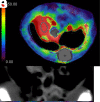
Figure 1.
Figure 1.
The 4D-CTP images of one rat in the 80% group at 1 h. Images A to D are axial HAP, PVP, HPI and CT plain images, respectively. Images E to H are coronal HAP, PVP, HPI and CT plain image, respectively. HAP, hepatic artery perfusion; HPI, hepatic perfusion index; PVP, portal vein perfusion.
Figure 2.
Figure 2.
HE staining of tissue samples from the sham group and 80% group. HE staining (×200 magnification). In the 80% group, obvious sinusoidal dilation appeared at 1 h post-operation; hepatocyte edema and vacuolar transformation appeared at 1 d and were more serious at 3 d; at 7 d, severe disruption of lobular architecture with extensive necrosis was detected. H&E,Haemotoxylin and Eosin.
Figure 3.
Figure 3.
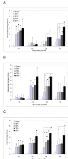
Morphological scores of the remnant livers of all groups. (A) Sinusoidal damage score. *p < 0.05, at 1 h, all of the PH groups vs the sham group; at 7 d, the 70 and 80% group vs the sham group; +p<0.05, the 80% group vs the 50 and 60% groups. (B) Hepatocyte injury score. *p < 0.05, at 1 d, all of the PH groups vs the sham group; at 3 d, the 60, 70 and 80% groups vs the sham group; +p<0.05, the 80% group vs the 50% group. (C) Liver damage score. *p < 0.05, at 1 h and 1 d, all of the PH groups vs the sham group; at 3 d, the 60, 70 and 80% groups vs the sham group; at 7 d, the 80% group vs the sham group. +p <0.05, at 3 d, the 80% group vs the 50 and 60% groups; at 7 d, the 80% group vs the 50% group. PH, partial hepatectomy.
Figure 4.
Figure 4.
Ki-67 staining of tissue samples from the sham group and 80% group. Ki-67 detection (×400 magnification). Hepatic nuclei are stainedbrownish yellow (Arrow). Compromised hepatocyte proliferation appeared at 1 din the 80% group, which was relieved at 3 d.
Figure 5.
Figure 5.
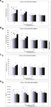
The CTP parameters in all of the PH groups. (A) PVP. *p < 0.05, at 1 h and 1 d, the 80% group vs the sham group;+p<0.05, at 1 h, the 80% group vs the 50 and 60% groups; (B) TLP.*p < 0.05, at 1 h and 1 d, the 80% group vs the sham group;+p<0.05, at 1 h, the 80% group vs the 50 and 60% groups; (C) HAP; (D) HPI. *p < 0.05, at 1 h and 1 d, the 80% group vs the sham group. CTP, CT perfusion; HAP, hepatic artery perfusion; HPI, hepatic perfusion index; PH, partial hepatectomy; PVP, portal vein perfusion; TLP, total liver perfusion.

Similar articles

References

    1. Yao S , Kaido T , Uozumi R , Yagi S , Miyachi Y , Fukumitsu K , et al. . Is portal venous pressure modulation still indicated for all recipients in living donor liver transplantation? Liver Transpl 2018. ; 24 : 1578 – 88 . doi: 10.1002/lt.25180 - DOI - PubMed
    1. Badawy A , Hamaguchi Y , Satoru S , Kaido T , Okajima H , Uemoto S . Evaluation of safety of concomitant splenectomy in living donor liver transplantation: a retrospective study . Transpl Int 2017. ; 30 : 914 – 23 . doi: 10.1111/tri.12985 - DOI - PubMed
    1. Scatton O , Cauchy F , Conti F , Perdigao F , Massault PP , Goumard C , et al. . Two-stage liver transplantation using auxiliary laparoscopically harvested grafts in adults: Emphasizing the concept of "hypersmall graft nursing" . Clin Res Hepatol Gastroenterol 2016. ; 40 : 571 – 4 . doi: 10.1016/j.clinre.2016.03.002 - DOI - PubMed
    1. Raut V , Alikhanov R , Belghiti J , Uemoto S . Review of the surgical approach to prevent small-for-size syndrome in recipients after left lobe adult LDLT . Surg Today 2014. ; 44 : 1189 – 96 . doi: 10.1007/s00595-013-0658-6 - DOI - PubMed
    1. Gyoten K , Mizuno S , Kato H , Murata Y , Tanemura A , Azumi Y , et al. . A novel predictor of posttransplant portal hypertension in Adult-To-Adult living donor liver transplantation: increased estimated Spleen/Graft volume ratio . Transplantation 2016. ; 100 : 2138 – 45 . doi: 10.1097/TP.0000000000001370 - DOI - PMC - PubMed