

Wnt3a involved in the mechanical loading on improvement of bone remodeling and angiogenesis in a postmenopausal osteoporosis mouse model
- PMID: 31017804
- PMCID: PMC9272758
- DOI: 10.1096/fj.201802711R
Wnt3a involved in the mechanical loading on improvement of bone remodeling and angiogenesis in a postmenopausal osteoporosis mouse model
Abstract
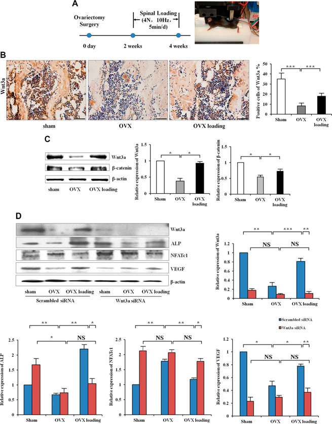
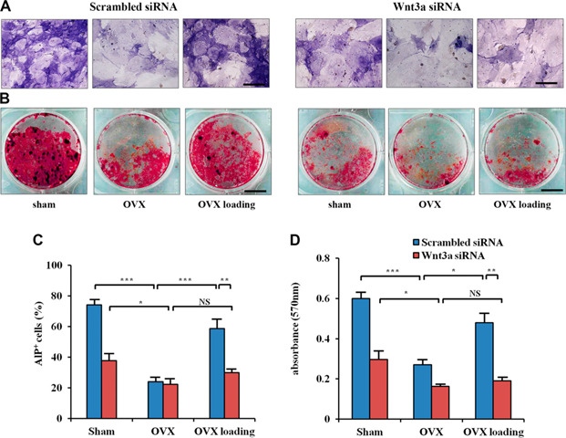
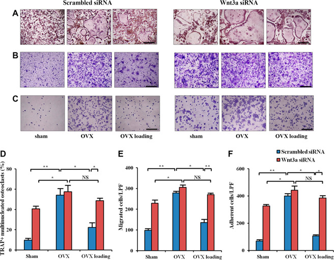
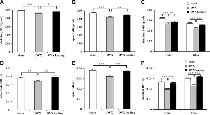
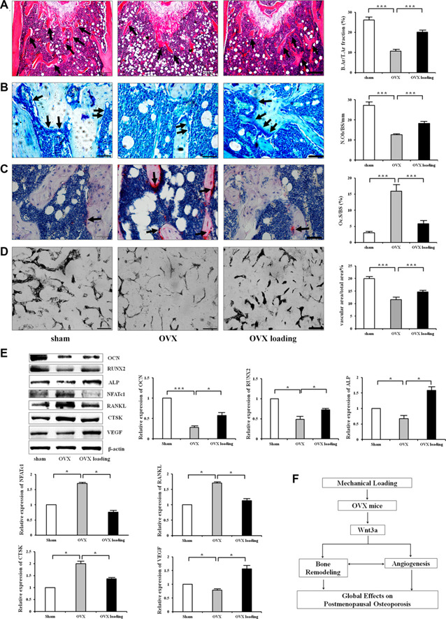
Osteoporosis is a major health problem, making bones fragile and susceptible to fracture. Previous works showed that mechanical loading stimulated bone formation and accelerated fracture healing. Focusing on the role of Wnt3a (wingless/integrated 3a), this study was aimed to assess effects of mechanical loading to the spine, using ovariectomized (OVX) mice as a model of osteoporosis. Two-week daily application of this novel loading (4 N, 10 Hz, 5 min/d) altered bone remodeling with an increase in Wnt3a. Spinal loading promoted osteoblast differentiation, endothelial progenitor cell migration, and tube formation and inhibited osteoclast formation, migration, and adhesion. A transient silencing of Wnt3a altered the observed loading effects. Spinal loading significantly increased bone mineral density, bone mineral content, and bone area per tissue area. The loaded OVX group showed a significant increase in the number of osteoblasts and reduction in osteoclast surface/bone surface. Though expression of osteoblastic genes was increased, the levels of osteoclastic genes were decreased by loading. Spinal loading elevated a microvascular volume as well as VEGF expression. Collectively, this study supports the notion that Wnt3a-mediated signaling involves in the effect of spinal loading on stimulating bone formation, inhibiting bone resorption, and promoting angiogenesis in OVX mice. It also suggests that Wnt3a might be a potential therapeutic target for osteoporosis treatment.-Li, X., Liu, D., Li, J., Yang, S., Xu, J., Yokota, H., Zhang, P. Wnt3a involved in the mechanical loading on improvement of bone remodeling and angiogenesis in a postmenopausal osteoporosis mouse model.
Keywords: OVX; bone formation; bone resorption; neovascularization; spinal load.
Figures

Similar articles
-
Piezoelectric Microvibration Mitigates Estrogen Loss-Induced Osteoporosis and Promotes Piezo1, MicroRNA-29a, and Wnt3a Signaling in Osteoblasts.Int J Mol Sci. 2021 Aug 31;22(17):9476. doi: 10.3390/ijms22179476. Int J Mol Sci. 2021. PMID: 34502380 Free PMC article.
-
Loading-driven PI3K/Akt signaling and erythropoiesis enhanced angiogenesis and osteogenesis in a postmenopausal osteoporosis mouse model.Bone. 2022 Apr;157:116346. doi: 10.1016/j.bone.2022.116346. Epub 2022 Feb 1. Bone. 2022. PMID: 35114427
-
Increased Osteoblastic Cxcl9 Contributes to the Uncoupled Bone Formation and Resorption in Postmenopausal Osteoporosis.Clin Interv Aging. 2020 Jul 20;15:1201-1212. doi: 10.2147/CIA.S254885. eCollection 2020. Clin Interv Aging. 2020. PMID: 32764906 Free PMC article.
-
Strontium ranelate in osteoporosis.Curr Pharm Des. 2002;8(21):1907-16. doi: 10.2174/1381612023393639. Curr Pharm Des. 2002. PMID: 12171530 Review.
-
Cell communication and relevant signaling pathways in osteogenesis-angiogenesis coupling.Bone Res. 2025 Apr 7;13(1):45. doi: 10.1038/s41413-025-00417-0. Bone Res. 2025. PMID: 40195313 Free PMC article. Review.
Cited by
-
Mechanical loading stimulates bone angiogenesis through enhancing type H vessel formation and downregulating exosomal miR-214-3p from bone marrow-derived mesenchymal stem cells.FASEB J. 2021 Jan;35(1):e21150. doi: 10.1096/fj.202001080RR. Epub 2020 Nov 8. FASEB J. 2021. PMID: 33161580 Free PMC article.
-
Vascularized Tissue Organoids.Bioengineering (Basel). 2023 Jan 17;10(2):124. doi: 10.3390/bioengineering10020124. Bioengineering (Basel). 2023. PMID: 36829618 Free PMC article. Review.
-
Mechanical Loading-Driven Tumor Suppression Is Mediated by Lrp5-Dependent and Independent Mechanisms.Cancers (Basel). 2021 Jan 13;13(2):267. doi: 10.3390/cancers13020267. Cancers (Basel). 2021. PMID: 33450808 Free PMC article.
-
Mechanical stimuli-mediated modulation of bone cell function-implications for bone remodeling and angiogenesis.Cell Tissue Res. 2021 Dec;386(3):445-454. doi: 10.1007/s00441-021-03532-6. Epub 2021 Oct 19. Cell Tissue Res. 2021. PMID: 34665321 Review.
-
Knee loading repairs osteoporotic osteoarthritis by relieving abnormal remodeling of subchondral bone via Wnt/β-catenin signaling.FASEB J. 2020 Feb;34(2):3399-3412. doi: 10.1096/fj.201902117R. Epub 2020 Jan 10. FASEB J. 2020. PMID: 31925860 Free PMC article.
References
-
- Deng, L. , Wang, Y. , Peng, Y. , Wu, Y. , Ding, Y. , Jiang, Y. , Shen, Z. , and Fu, Q. (2015) Osteoblast‐derived microvesicles: a novel mechanism for communication between osteoblasts and osteoclasts. Bone 79, 37–42 - PubMed
-
- Xu, B. , Lovre, D. , and Mauvais‐Jarvis, F. (2016) Effect of selective estrogen receptor modulators on metabolic homeostasis. Biochimie 124, 92–97 - PubMed
Publication types
MeSH terms
Substances
Grants and funding
LinkOut - more resources
Full Text Sources
