Prolonged time course of population excitatory postsynaptic potentials in motoneurons of chronic stroke survivors
- PMID: 31017842
- PMCID: PMC6689790
- DOI: 10.1152/jn.00288.2018
Prolonged time course of population excitatory postsynaptic potentials in motoneurons of chronic stroke survivors
Abstract
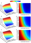
Hyperexcitability of spinal motoneurons may contribute to muscular hypertonia after hemispheric stroke. The origins of this hyperexcitability are not clear, but we hypothesized that prolongation of the Ia excitatory postsynaptic potential (EPSP) in spastic motoneurons may be one potential mechanism, by enabling more effective temporal summation of Ia EPSPs, making action potential initiation easier. Thus, the purpose of this study is to quantify the time course of putative EPSPs in spinal motoneurons of chronic stroke survivors. To estimate the EPSP time course, a pair of low-intensity electrical stimuli was delivered sequentially to the median nerve in seven hemispheric stroke survivors and in six intact individuals, to induce an H-reflex response from the flexor carpi radialis muscle. H-reflex response probability was then used to quantify the time course of the underlying EPSPs in the motoneuron pool. A population EPSP estimate was then derived, based on the probability of evoking an H-reflex from the second test stimulus in the absence of a reflex response to the first conditioning stimulus. Our experimental results showed that in six of seven hemispheric stroke survivors, the apparent rate of decay of the population EPSP was markedly slower in spastic compared with contralateral (stroke) and intact motoneuron pools. There was no significant difference in EPSP time course between the contralateral side of stroke survivors and control subject muscles. We propose that one potential mechanism for hyperexcitability of spastic motoneurons in chronic stroke survivors may be associated with this prolongation of the Ia EPSP time course. Our subthreshold double-stimulation approach could provide a noninvasive tool for quantifying the time course of EPSPs in both healthy and pathological conditions. NEW & NOTEWORTHY Spastic motoneurons in stroke survivors showed a prolonged Ia excitatory postsynaptic potential (EPSP) time course compared with contralateral and intact motoneurons, suggesting that one potential mechanism for hyperexcitability of spastic motoneurons in chronic stroke survivors may be associated with this prolongation of the Ia EPSP time course.
Keywords: EPSP time course; double stimulation; reflex; spasticity; stroke.
Conflict of interest statement
No conflicts of interest, financial or otherwise, are declared by the author(s).
Figures


Similar articles
-
Estimating the time course of population excitatory postsynaptic potentials in motoneurons of spastic stroke survivors.J Neurophysiol. 2015 Mar 15;113(6):1952-7. doi: 10.1152/jn.00946.2014. Epub 2014 Dec 24. J Neurophysiol. 2015. PMID: 25540228 Free PMC article.
-
Excitatory synaptic potentials in spastic human motoneurons have a short rise-time.Muscle Nerve. 2005 Jul;32(1):99-103. doi: 10.1002/mus.20318. Muscle Nerve. 2005. PMID: 15786417
-
Effects of chronic spinalization on ankle extensor motoneurons. I. Composite monosynaptic Ia EPSPs in four motoneuron pools.J Neurophysiol. 1994 Apr;71(4):1452-67. doi: 10.1152/jn.1994.71.4.1452. J Neurophysiol. 1994. PMID: 8035227
-
Partitioning of monosynaptic Ia excitatory postsynaptic potentials in the motor nucleus of the cat lateral gastrocnemius muscle.J Neurophysiol. 1986 Mar;55(3):569-86. doi: 10.1152/jn.1986.55.3.569. J Neurophysiol. 1986. PMID: 3514815 Review.
-
Synaptic mechanisms and circuitry involved in motoneuron control during sleep.Int Rev Neurobiol. 1983;24:213-58. doi: 10.1016/s0074-7742(08)60223-8. Int Rev Neurobiol. 1983. PMID: 6197386 Review.
Cited by
-
Research progress in extracorporeal shock wave therapy for upper limb spasticity after stroke.Front Neurol. 2023 Feb 9;14:1121026. doi: 10.3389/fneur.2023.1121026. eCollection 2023. Front Neurol. 2023. PMID: 36846123 Free PMC article. Review.
References
Publication types
MeSH terms
Grants and funding
LinkOut - more resources
Full Text Sources
Medical

