. 2019 Apr 24;15(4):e1008034.
doi: 10.1371/journal.pgen.1008034. eCollection 2019 Apr.

CD34 defines melanocyte stem cell subpopulations with distinct regenerative properties

Affiliations

CD34 defines melanocyte stem cell subpopulations with distinct regenerative properties

Sandeep S Joshi et al. PLoS Genet. .

Abstract

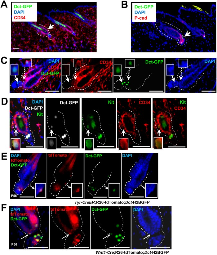
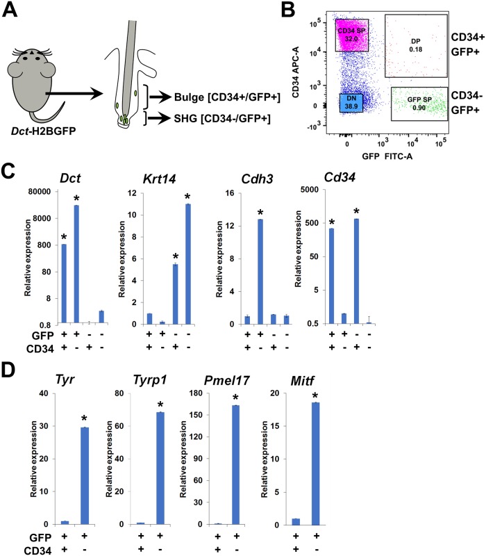
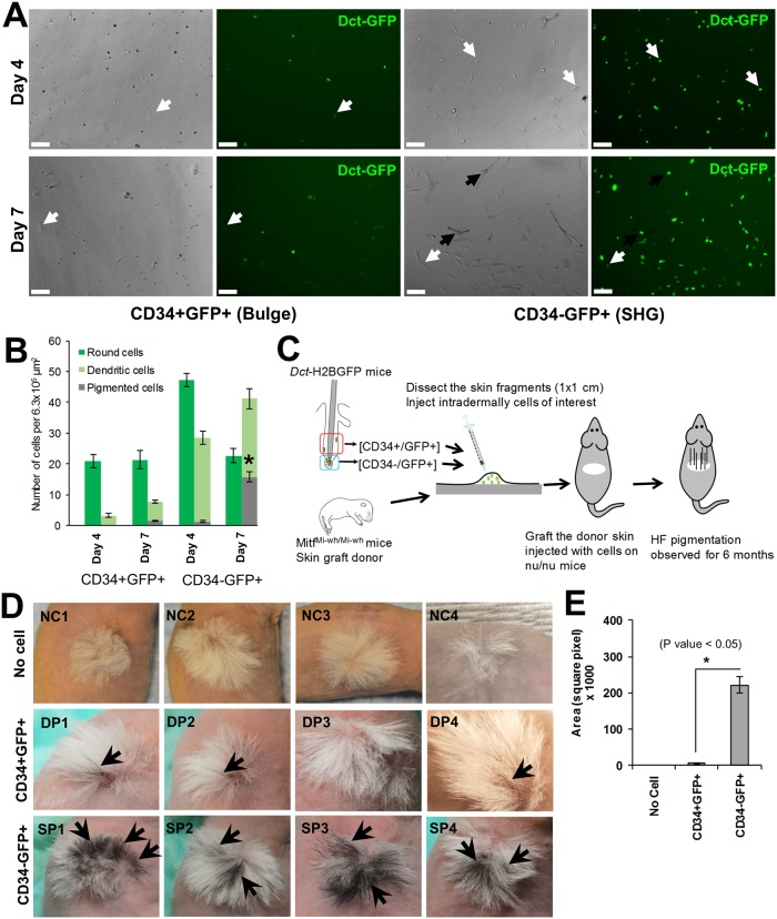
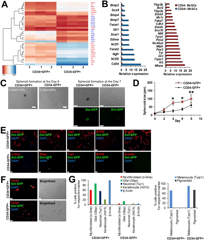
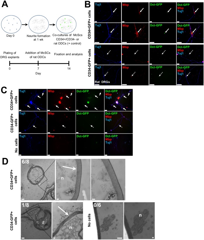
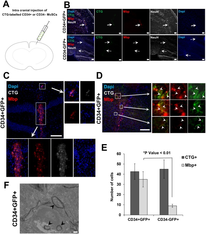
Melanocyte stem cells (McSCs) are the undifferentiated melanocytic cells of the mammalian hair follicle (HF) responsible for recurrent generation of a large number of differentiated melanocytes during each HF cycle. HF McSCs reside in both the CD34+ bulge/lower permanent portion (LPP) and the CD34- secondary hair germ (SHG) regions of the HF during telogen. Using Dct-H2BGFP mice, we separate bulge/LPP and SHG McSCs using FACS with GFP and anti-CD34 to show that these two subsets of McSCs are functionally distinct. Genome-wide expression profiling results support the distinct nature of these populations, with CD34- McSCs exhibiting higher expression of melanocyte differentiation genes and with CD34+ McSCs demonstrating a profile more consistent with a neural crest stem cell. In culture and in vivo, CD34- McSCs regenerate pigmentation more efficiently whereas CD34+ McSCs selectively exhibit the ability to myelinate neurons. CD34+ McSCs, and their counterparts in human skin, may be useful for myelinating neurons in vivo, leading to new therapeutic opportunities for demyelinating diseases and traumatic nerve injury.


Conflict of interest statement

The authors declare no competing financial interests.

Figures

Fig 1
Fig 1. Identification of GFP-expressing melanocyte precursor cells in bulge and SHG of telogen HF.
Distinct subpopulations of GFP-expressing cells in the CD34+ bulge region (arrows, A) and P-Cad+ SHG region (arrows, B) in P56 dorsal skin HFs. Scale bars: (A) 100 μm (B) 50 μm. (C) GFP-expressing cells and anti-CD34 immunofluorescence in P56 dorsal skin HF. Dotted arrow depicts co-localization of bulge GFP-expressing cell and CD34 expression and solid arrow shows SHG GFP-expressing cells lack CD34 expression. Scale bars: 50 μm. (D) Immunofluorescence of Kit and CD34 expression in bulge GFP-expressing cells in P56 dorsal skin HF. Arrow depicts co-localization of bulge GFP-expressing cell with Kit and CD34 expression. Scale bars: 25 μm. (E & F) Co-localization of Tyr (E) or Wnt1 (F) driven tdTomato expression and GFP-expressing cells from both bulge and SHG (arrows) in telogen HF of Tyr-CreER;R26-tdTomato;Dct-H2BGFP or Wnt1-Cre;R26-tdTomato;Dct-H2BGFP mice at P56 respectively. Scale bars: 50 μm.
Fig 2
Fig 2. Separation of bulge and SHG GFP-expressing melanocyte precursor cells of telogen HFs.
(A) Experimental scheme: McSCs identified in HF bulge and SHG of Dct-H2BGFP mice were separated using FACS with GFP and anti-CD34. (B) Separation of bulge (CD34+GFP+) and SHG (CD34-GFP+) melanocyte precursor cells and CD34+GFP- and CD34-GFP- dermal cells using FACS with GFP and anti-CD34, showing a representative image of the FACS. DP = Double positive, SP = Single positive and DN = Double negative. (C) Quantitative RT-PCR analysis for the expression of Dct, Krt14, Cdh3, and Cd34 genes among the CD34+GFP+(bulge), CD34-GFP+(SHG), CD34+GFP- and CD34-GFP- sorted cell populations. Here, Gapdh was used as reference gene, (*P ≤ 0.01 by ANOVA). (D) Quantitative RT-PCR analysis for the expression of Tyr, Tyrp1 and Pmel17 in bulge (CD34+GFP+) and SHG (CD34+GFP+) sorted cells. Here, Gapdh was used as reference gene. (*P ≤ 0.01 by ANOVA).
Fig 3
Fig 3. Distinct melanogenic properties of bulge and SHG melanocyte precursor cells of telogen HFs.
(A) In vitro melanocyte differentiation potential of CD34+GFP+ and CD34-GFP+ McSCs in melanocyte differentiation culture condition for day 4 (top two panels) or day 7 (bottom two panels). Dark arrow indicates pigmented cells and white arrow indicates dendritic but non-pigmented cells. Scale bars: 100 μm. (B) Quantification of bulge and SHG McSCs potential to produce pigmented melanocytes in melanocyte differentiation medium at day 4 and 7. *P ≤ 0.01. (C) Viable CD34+GFP+ and CD34-GFP+ McSCs isolated from Dct-H2BGFP mice were injected into skin fragments of neonatal MitfMi-wh/Mi-wh mice (P0 –P2). Injected skin fragments were engrafted onto nude mice to observe regeneration of HF pigmentation. (D) Skin grafts following reconstitution with CD34-GFP+ cells (SP) showed significantly greater regeneration of follicular pigmentation than grafts receiving CD34+GFP+ cells (DP). HF pigmented regions of the grafted skin are indicated by arrows. No cell (NC) injected grafted skin fragments were used as a negative control and showed no pigmented HFs. (E) Quantification of pigmented region of engrafted skin receiving CD34- McSCs, or CD34+ McSCs, or no cells. Area of pigmented region was calculated using Image J software. (* P Value < 0.05. by ANOVA; N = 5).
Fig 4
Fig 4. CD34+ bulge McSCs exhibit distinct neural crest lineage potential.
(A) Heatmap of scaled and clustered, variance stabilizing transformation (VST) read count values obtained for select genes from RNA-seq analysis of CD34+ and CD34- McSCs. In bulge and SHG McSCs, the melanogenic marker (red) and NCSC marker (blue) are differentially expressed, closely clustered and are statistically significant. (Padjusted < 0.02, Benjamini-Hochberg adjusted p value) (B) RNA-Seq analysis, based on fold change, showed that CD34+GFP+ McSCs (blue; left panel) express higher NCSC markers, whereas CD34-GFP+ McSCs (red; right panel) express higher melanogenic markers. (C) Formation of non-adherent spheroids was studied among CD34+GFP+ (bulge) and CD34-GFP+ (SHG) McSCs when cultured in NCSC medium (top panels). The image in bottom panel depicts retention of GFP expression in spheroids formed by both cell types. The top panel also shows the percentage of cells initially plated that formed spheroids from three independent experiments. (N = 3, standard deviation reported) (D) The size of non-adherent spheroids derived from bulge and SHG McSCs when cultured in NCSC medium as determined at days 2, 4, 6 and 8. (*P ≤ 0.01, **P Value ≤ 0.05 by ANOVA; N = 5). (E) Expression of neural crest lineage markers Gfap, α-Sma, Tuj1, Krt15 among CD34+GFP+ (top panel) and CD34-GFP+ (bottom panel) McSCs following adherent culture in neural crest differentiation medium. Scale bars: 50 μm. (F) Expression of melanocyte lineage marker Tyrp1 among CD34+GFP+ (top panel) and CD34-GFP+ (bottom panel) McSCs following adherent culture in melanocyte differentiation medium. In right two panels, brightfield images show pigmented melanocytes among cultured CD34+GFP+ (top) and CD34-GFP+ (bottom) McSCs. Scale bars: 50 μm. (G) Quantification of neural crest-derived cell and melanocyte marker expression frequency after cells were cultured in either neural crest differentiation (left) or melanocyte differentiation (right) condition. For this experiment, all cells in each well were counted and this graph is a representative of 3 independent experiments.
Fig 5
Fig 5. Myelination properties of CD34+ bulge McSCs.
(A) Schematic of in vitro DRG co-culture system: CD34+, CD34- McSCs or rat ODCs and DRG cells (isolated from rat E17 or P5 shi/shi mice) were co-cultured for one week and screened for myelination of axons by immunofluorescence and by EM. Bottom panel shows a timeline of culturing DRG explants at Day 0, addition of McSCs (experimental) and rat ODCs (positive control) at Day 7 and fixation of cells and analysis on day 14. (B & C) Co-cultures of CD34+ or CD34- McSCs and rat eDRGs (B), or neonatal shi/shi DRGs (C). Arrows point to GFP-expressing cells, with solid arrow representing CD34+ bulge McSCs and dotted arrow representing CD34- SHG McSCs. In (B), second and fourth rows represents high magnification images of first and third row images. Scale bars: 50 μm. (D) Electron-dense myelin sheath formation around shi/shi neurites when co-cultured with CD34+ or CD34- McSCs or no added cells using EM. In the right panels in either CD34+ or CD34- McSCs co-cultured with shi/shi DRGs, high magnification images of the region marked with black box are shown. In the no added cells sample images with shi/shi DRGs, right image is a high magnification of the left image. Arrow points to the dense myelin sheath and “n” indicates neurites. Scale bars: 500 nm (non-magnified images) and 100 nm (high magnified images).
Fig 6
Fig 6. In vivo glial differentiation potential of CD34+ bulge McSCs.
(A) Schematic diagram depicting transplantation of CD34+ or CD34- McSCs, after labelling with fluorescent CTG dye, intracranially into shi/shi mouse brain. At the end of the experiment, shi/shi mice brains were assessed for the presence of CTG cells at 2 weeks post-injection. (B) Cranial sections of shi/shi brains transplanted with CD34+ or CD34- McSCs show co-localization of Mbp expression in CTG-labelled CD34+ McSCs (solid arrows); co-localization is absent following transplantation of CTG-labelled CD34- McSCs (dotted arrows). (C) Cranial section of shi/shi brain transplanted with CD34+ McSCs shows individual CTG-labelled CD34+ McSCs co-expressing Mbp (top and bottom inset boxes). (D) A section of myelin-deficient shi/shi mice brain receiving CD34+ McSCs depicts co-localization of Mbp with neurofilament-H stained neurons in the surrounding vicinity of transplanted CTG-labelled CD34+ McSCs (arrowhead pointing within inset boxes). Scale bars: 100 μm. (E) Quantification of co-localization of Mbp expression in CTG-labelled CD34+ McSCs in cranial sections of shi/shi brains transplanted with CD34+ or CD34- McSCs. (*P ≤ 0.01 analyzed by ANOVA; N = 5) (F) Representative example of dense myelin sheath analyzed by transmission electron microscopy, surrounding neuron of CD34+ McSC-transplanted shi/shi brain (arrowhead). Scale bars: 500 nm.

References

    1. Osawa M, Egawa G, Mak SS, Moriyama M, Freter R, et al. (2005) Molecular characterization of melanocyte stem cells in their niche. Development 132(24): 5589–5599. 10.1242/dev.02161 - DOI - PubMed
    1. Nishimura EK, Jordan SA, Oshima H, Yoshida H, Osawa M, et al. (2002) Dominant role of the niche in melanocyte stem-cell fate determination. Nature 416(6883): 854–860. 10.1038/416854a - DOI - PubMed
    1. Cotsarelis G. (2006) Epithelial stem cells: A folliculocentric view. J Invest Dermatol 126(7): 1459–1468. 10.1038/sj.jid.5700376 - DOI - PubMed
    1. Schneider MR, Schmidt-Ullrich R, Paus R. (2009) The hair follicle as a dynamic miniorgan. Curr Biol 19(3): R132–42. 10.1016/j.cub.2008.12.005 - DOI - PubMed
    1. Greco V, Chen T, Rendl M, Schober M, Pasolli HA, et al. (2009) A two-step mechanism for stem cell activation during hair regeneration. Cell Stem Cell 4(2): 155–169. 10.1016/j.stem.2008.12.009 - DOI - PMC - PubMed

Publication types

MeSH terms