Review
. 2020;12(1):21-30.
doi: 10.1159/000497414. Epub 2019 Apr 24.

Autophagy in Pulmonary Innate Immunity

Affiliations
Review

Autophagy in Pulmonary Innate Immunity

Lang Rao et al. J Innate Immun. 2020.

Abstract

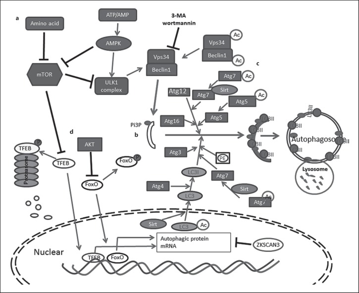
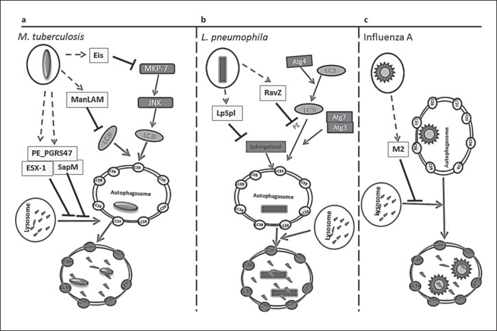
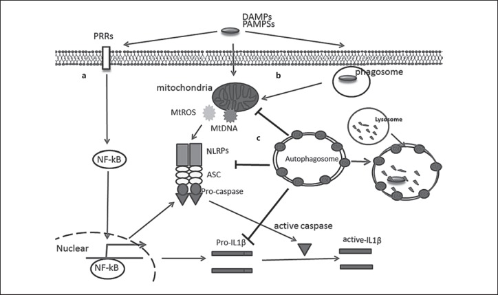
Autophagy is a major intracellular digestion system that delivers cytoplasmic components for degradation and recycling. In this capacity, autophagy plays an important role in maintaining cellular homeostasis by mediating the degradation of cellular macromolecules and dysfunctional organelles and regeneration of nutrients for cell growth. Autophagy is important in innate immunity, as it is responsible for the clearance of various pathogens. Deficiency of intracellular autophagy can result in exaggerated activation of the inflammasome. The latter is an innate immune complex that senses diverse pathogen-associated or danger-associated molecular patterns and activates the expression of inflammatory cytokines. In autophagy-deficient cells, accumulation of damaged organelles, misfolded proteins, and reactive oxygen species contribute to inflammasome activation. The lung is continuously exposed to pathogens from the environment, rendering it vulnerable to infection. The lung innate immune cells act as a crucial initial barrier against the continuous threat from pathogens. In this review, we will summarize recent findings on the regulation of autophagy and its inter-action with innate immunity, focusing on the lung.

Keywords: Autophagy; Inflammasome; Innate immunity and pulmonary inflammation; Pathogen.


Conflict of interest statement

The authors have no conflicts of interest to declare.

Figures

Fig. 1
Fig. 1
Simplified autophagy signaling pathway and regulation. a Nutrition deprivation inhibits mTOR, which in turn activates ULK1 complex. Energy deprivation is sensed by AMPK to suppress mTOR and activates ULK1 complex. Activated ULK1 complex recruits Vps34-Beclin-1 complex to produce PI3P and initiates phagosome formation. b Two conjugation systems, Atg12–Atg5-Atg16L and LC3–PE, facilitate phagophore membrane elongation and autophagosome maturation. c NAD-dependent deacetylase Sirt1 catalyzes deacetylation of Vps34, Beclin1, Atg5, Atg7, and LC3 to promote autophagy. d Transcription factors TFEB and FoxO translocate to the nuclear to induce autophagy-related gene expression. Their transcription activity can be inhibited by phosphorylation caused by mTOR or AKT, respectively. Phosphorylated TFEB is digested by proteasome; ZKSCAN3 repress autophagy-related gene transcription.
Fig. 2
Fig. 2
Strategies used by pulmonary pathogens to avoid host autophagy. a M. tuberculosis has five identified anti-autophagy factors. M. tuberculosis secretion system Esx-1, the secreted phosphatase SapM, and the virulence protein PE_PGRS47 inhibit autophagy by blocking autophagosome lysosome fusing. Eis is an N-acetyltransferase. It acetylates JNK-specific phosphatase MKP-7 to initiate the inhibition of JNK-dependent autophagy. Mannose-capped lipoarabinomannan ManLAM interferes with trafficking proteins in autophagy and also affects LC3 protein expression levels and inhibits accumulation of autophagic vacuoles. b RavZ, the bacterial effector protein of L. pneumophila, inhibits autophagy by hydrolyzing the release of LC3II phosphatidylethanolamine at the carboxyl-terminal glycine residue. The hydrolyzed LC3 cannot be reconjugated by Atg7 and Atg3 to form mature LC3II for autophagosome localization. LpSpl with sphingosine-1 phosphate lyase activity reduces sphingolipid levels, reducing ­autophagosome formation. c Influenza A's transmembrane protein M2, which arrests autophagosome degradation by blocking autophagosome and lysosome fusion.
Fig. 3
Fig. 3
Autophagy in host defense and inflammasome regulation. a PAMPs or DAMPs, recognized by pattern-recognition receptors, result in NF-κB activation. Active NK-κB promotes inflammasome components and cytokine expression. b PAMPs or DAMPs cause mitochondrial damage and the release of mitochondrial ROS and DNA, triggering the assembling of NLRP3, ASC, and Pro-caspase into active inflammasome. Caspase-1 is activated by autocleavage and then cleaves the pro-inflammatory cytokines IL-1β into active cytokines. Bacteria-containing phagosome membrane disruption leads to the release of PAMPs. c Activated auto­phagosomes can engulf damaged mitochondria, NLRP3, ASC, and Pro-caspase, and target them to lysosomal degradation, reducing the production and secretion of active cytokines.

References

    1. Harnett MM, Pineda MA, Latré de Laté P, Eason RJ, Besteiro S, Harnett W, et al. From Christian de Duve to Yoshinori Ohsumi: more to autophagy than just dining at home. Biomed J. 2017 Feb;40((1)):9–22. - PMC - PubMed
    1. Klionsky DJ, Emr SD. Autophagy as a regulated pathway of cellular degradation. Science. 2000 Dec;290((5497)):1717–21. - PMC - PubMed
    1. Mizushima N, Komatsu M. Autophagy: renovation of cells and tissues. Cell. 2011 Nov;147((4)):728–41. - PubMed
    1. Gutierrez MG, Master SS, Singh SB, Taylor GA, Colombo MI, Deretic V. Autophagy is a defense mechanism inhibiting BCG and Mycobacterium tuberculosis survival in infected macrophages. Cell. 2004 Dec;119((6)):753–66. - PubMed
    1. Jagannath C, Lindsey DR, Dhandayuthapani S, Xu Y, Hunter RL, Jr, Eissa NT. Autophagy enhances the efficacy of BCG vaccine by increasing peptide presentation in mouse dendritic cells. Nat Med. 2009 Mar;15((3)):267–76. - PubMed