Review
. 2019 Apr 23;20(8):1996.
doi: 10.3390/ijms20081996.

Two Sides of the Coin: Ezrin/Radixin/Moesin and Merlin Control Membrane Structure and Contact Inhibition

Affiliations
Review

Two Sides of the Coin: Ezrin/Radixin/Moesin and Merlin Control Membrane Structure and Contact Inhibition

Katharine A Michie et al. Int J Mol Sci. .

Abstract

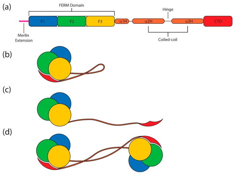
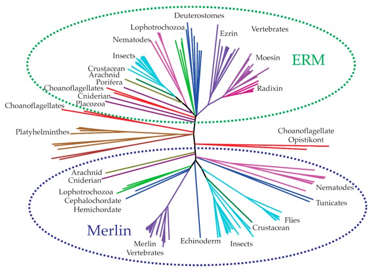
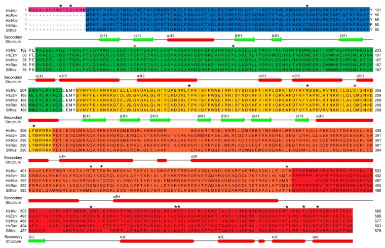
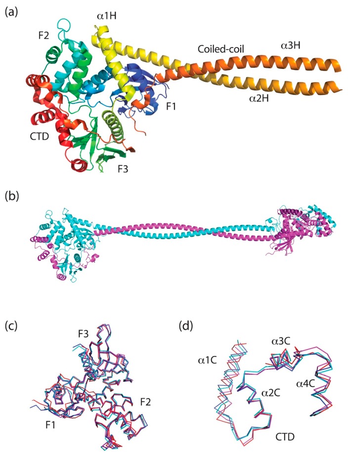
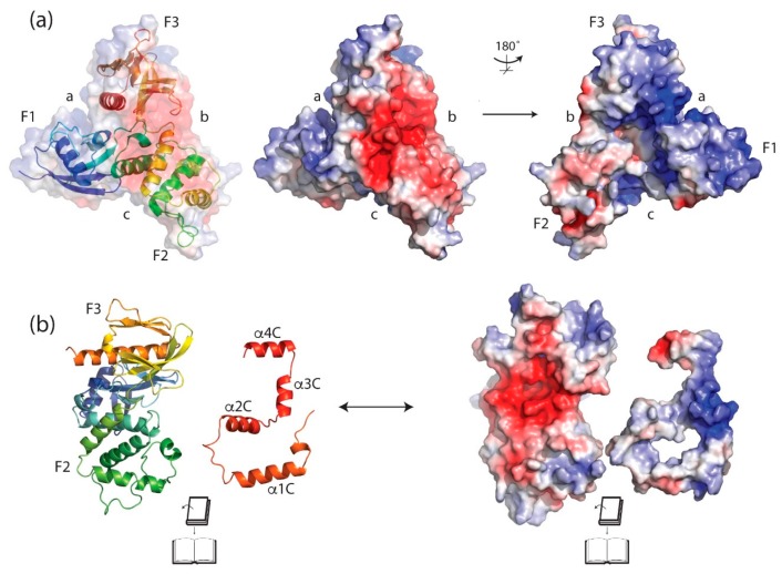
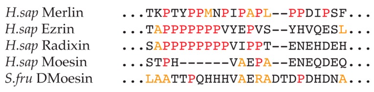
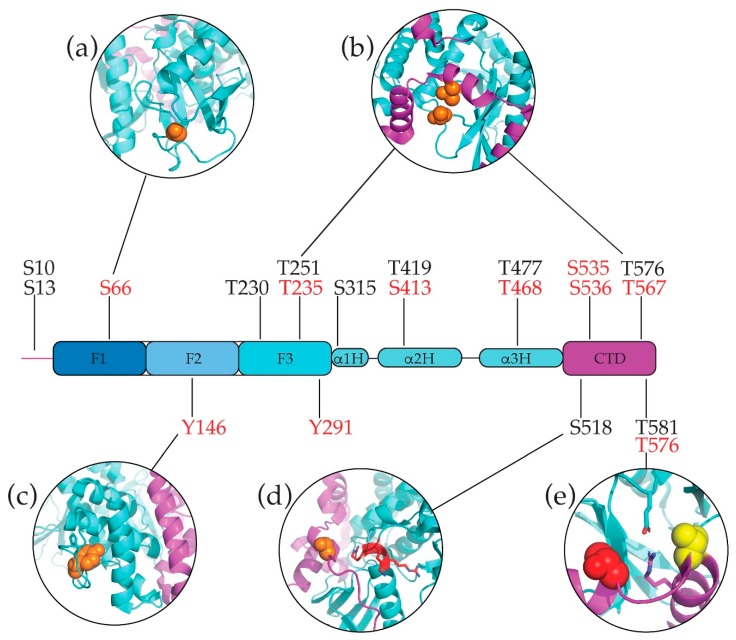
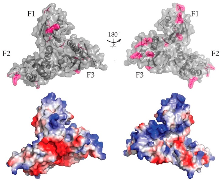
The merlin-ERM (ezrin, radixin, moesin) family of proteins plays a central role in linking the cellular membranes to the cortical actin cytoskeleton. Merlin regulates contact inhibition and is an integral part of cell-cell junctions, while ERM proteins, ezrin, radixin and moesin, assist in the formation and maintenance of specialized plasma membrane structures and membrane vesicle structures. These two protein families share a common evolutionary history, having arisen and separated via gene duplication near the origin of metazoa. During approximately 0.5 billion years of evolution, the merlin and ERM family proteins have maintained both sequence and structural conservation to an extraordinary level. Comparing crystal structures of merlin-ERM proteins and their complexes, a picture emerges of the merlin-ERM proteins acting as switchable interaction hubs, assembling protein complexes on cellular membranes and linking them to the actin cytoskeleton. Given the high level of structural conservation between the merlin and ERM family proteins we speculate that they may function together.

Keywords: FERM domain; ezrin; merlin; moesin; radixin.


Conflict of interest statement

The authors declare no conflict of interest.

Figures

Figure 1
Figure 1
Schematic overview of merlin-ERM (ezrin, radixin and moesin) protein domain structure and states. (a) The conserved domain structure of merlin-ERM proteins. The N-terminal FERM (band 4.1 protein, ezrin, radixin, moesin) domain comprises three subdomains, F1 (blue), F2 (green) and F3 (yellow). This is followed by a central helical domain comprising three α helices (orange), with the latter two, α2H and α3H, forming a coiled-coil in the monomer structure. At the C-terminus lies the largely α helical C-terminal domain (CTD, red). Note that merlin contains an N-terminal extension that is not seen in the ERM proteins (magenta). (bd) show various states of merlin-ERM proteins. (b) represents the closed state monomer structure where the CTD and FERM domains form a globular structure with the α helical coiled-coil protruding. (c) represents the putative open state, where an extended “helical” domain separates the FERM and CTD domains. (d) shows the domain-swapped dimer state.
Figure 2
Figure 2
Phylogenetic tree demonstrating the divergence of merlin from the ERM proteins. The upper arbor shows ERM proteins spanning early branching metazoa through to vertebrates. The vertebrate ERMs diverge into distinct ezrin, radixin and moesin clades (top right). The lower arbor shows merlin proteins again spanning early branching metazoa through to vertebrates. Near the center of the tree lie various merlin-ERM proteins where it is not clear that they can be classified as either merlin or ERM from sequence analysis alone. We note that there are multiple choanoflagellate proteins (red branches) that appear to represent distinct merlin and ERM proteins. Sequences were obtained from the National Center for Biotechnology Information (NCBI, US National Library of Medicine) non-redundant protein database using the program BLAST [14]. Multiple sequence alignment and neighbor-joining phylogenetic tree were constructed with the program MUSCLE [15] using the EMBL-EBI webserver. The tree was drawn using the program FigTree v1.4.2 [19].
Figure 3
Figure 3
Multiple sequence alignment of human merlin-ERMs plus Sfmoesin. Domains are colored as per Figure 1 (FERM subdomain F1—blue, FERM F2—green, FERM F3—yellow, Helical domain—orange, CTD—red). Secondary structural elements are shown based on the Sfmoesin crystal structure. Black stars denote phosphorylation sites discussed in the text. Red stars indicate the two conserved cysteine residues. Sequence alignment was carried out with the program MUSCLE [15] using the EMBL-EBI webserver. The figure was prepared using the program Jalview [21].
Figure 4
Figure 4
The structure of merlin-ERM. (a) The crystal structure of the only full-length ERM, Sfmoesin, from Spodoptera frugiperda (PDB 2I1K) [55]. The protein is colored as a rainbow from the blue N-terminus to the red C-terminus. A striking feature is the coiled-coil extending from the globular FERM:CTD complex. (b) Model of full-length human ezrin domain-swapped homo-dimer which is based on the crystal structure of the ezrin FERM:CTD complex plus small-angle x-ray scattering data [56]. (c) Wire overlay of FERM domains (from crystal structures of FERM:CTD complexes of human merlin (4ZRJ, red) [62], ezrin (4RM9, Blue) [56] and moesin (1EF1, cyan) [61] and S. frugiperda ERM (2I1K, magenta) [55] displaying almost identical tertiary structure. (d) Wire overlay of the final α helical section of the CTD taken from crystal structures of FERM:CTD complexes (same structures as (c)), further displaying the tertiary similarity. This figure was made using the program PyMOL [63].
Figure 5
Figure 5
Surface electrostatics of the human ezrin. (a) Surface electrostatics of the ezrin FERM domain (PDB 4RMA, [56]). Left panel shows the protein backbone (ribbon diagram) overlayed with a transparent electrostatic surface. The clefts between FERM subdomains are labeled a, b and c. Middle panel shows the electrostatic surface in the same view, while the right panel is rotated 180° about a vertical axis in the plane of the page. The cleft between FERM subdomains F2 and F3 (labeled b) shows a large area of negative charge (red, middle panel) while the cleft between subdomains F1 and F3 (labeled a) shows a large surface of positive charge (blue, right panel). (b) The interface between the FERM and CTD complex is visualized by separating these domains and rotating the CTD by 180° about a vertical axis in the plane of the page (left panel). The right panel shows the electrostatic surface potential calculated for the separated FERM and CTD domains. It is clear that charge complementarity is important in stabilizing the FERM:CTD complex. Image rendered in PyMOL [63] showing Poisson–Boltzmann electrostatic potential calculated with PDB2PQR [66].
Figure 6
Figure 6
ClustalOmega sequence alignment of the proline-rich/polyproline region at the start of the CTD (just after the end of the coiled-coil in the helical domain). The alignment contains human merlin-ERM proteins and the insect ERM (Sfmoesin) from S. frugiperda. Prolines highlighted in red and other residues more commonly found in Type II poly-proline helices highlighted in orange. The alignment was carried out using Clustal Omega [80].
Figure 7
Figure 7
Montage of merlin-ERM proteins interacting with peptides mimicking binding partner proteins. Left panel shows the Sfmoesin crystal structure [55] as a reference with domains colored as per Figure 1. On the right, individual panels show FERM:peptide complexes with the FERM colored as per Figure 1 and the peptides shown in magenta. The complexes are: moesin:Crumbs (PDB accession 4YL8 [107]); radixin:CD44 (2ZPY [104]); radixin:MT1-MMP (3X23 [106]); radixin:EBP50 (2D10 [108]); merlin:Lats1 (4ZRK [62]).; and merlin:DCAF1 (4P7I [111]). We note that for all complexes, with the exception of the radixin:MT1-MMP complex, the structure of the bound peptide mimics a portion of the CTD domain in the Sfmoesin structure (compare individual complex panels with the Sfmoesin structure). All structures are oriented the same way for direct comparison. The panels were rendered in PyMOL [63].
Figure 8
Figure 8
Structural landscape of some of merlin-ERM’s phosphorylation sites which are represented as atom spheres. (a) ezrin Ser66, a threonine in moesin and radixin and not conserved in merlin. This residue is located in subdomain F1 in the long loop between β strands β4F1 and β5F1. (b) Two conserved threonines, one of which (merlin Thr251, ezrin Thr235; lower one in panel) lies in a conserved loop between β strands β3F3 and β4F3 in subdomain F3, while the other is located on α3C (merlin Thr576, ezrin Thr567; upper one in panel). They are directly opposite each other and phosphorylation of both would likely destabilize the FERM–CTD interaction. (c) ERM Tyr146 that faces into the center of subdomain F2. In merlin and Sfmoesin this residue is a phenylalanine, preserving the aromaticity. (d) When mapped onto the full-length Sfmoesin structure, merlin Ser518 lies directly opposite one of the most conserved regions in all merlin-ERM proteins: 308MRRRK312 at the C-terminus of α1F3 in subdomain F3 (shown in red) which is highly positively charged. (e) Conserved ERM threonine (ezrin Thr576 but Ala585 in merlin; yellow) and merlin Thr581 (but conserved ERM arginine, ezrin Arg572, red). The two phosphorylatable threonines are proximal to the highly conserved and functionally important salt bridge (merlin Glu260–Arg588; ezrin Glu244–Arg579; Section 4.4). The panels were rendered in PyMOL [63].
Figure 9
Figure 9
Ubiquitination and acetylation sites (pink) mapped onto the crystal structure of ezrin FERM domain (surface rendered in grey), shown in two orientations related by 180° rotation about a vertical axis in the plane of the page (top two panels). The two lower panels show the local electrostatic potential surfaces for the same two orientations of ezrin FERM when it is unmodified (i.e., not ubiquitinated nor acetylated). The coloring scheme is red for negative electrostatic potential through to blue for positive electrostatic potential. Image rendered in PyMOL [63] showing Poisson–Boltzmann electrostatic potential calculated with PDB2PQR [66].
Figure 10
Figure 10
Structural insights into merlin-ERM lipid binding. (a) Structure of the full-length Sfmoesin (PDB: 2I1K) indicating the three proposed lipid binding sites; the Patch (green), the Pocket (magenta) and the Pouch (cyan). The FERM domain is shown in grey, with the helical domain orange and the CTD in red. (b) The lipid-binding sites on the Sfmoesin FERM domain in the same orientation and coloring as per (a). (c) Sequence conservation of the lipid-binding sites. Coloring for the different lipid-binding sites are consistent across panels (ac). (d) and (e) Electrostatic maps of the full-length Sfmoesin and the Sfmoesin FERM domain, respectively. The coloring scheme is red for negative electrostatic potential through to blue for positive electrostatic potential. The orientation of these structures is consistent with panels (a) and (b). (f) Structural overlay of the Sfmoesin FERM domain (grey) with the radixin FERM (blue) bound to IP3 (sticks representation) in the Pocket (PDB: 1GC6). (g) Structural overlay of the Sfmoesin FERM (grey) with merlin FERM (gold) bound to a short chain PI(4,5)P2 (sticks representation) in the Pouch (PDB: 6CDS). The overlays in Panels (f) and (g) are in the same orientation as Sfmoesin FERM in Panel (b). (h) Orientation of the full-length Sfmoesin monomer structure docked onto a lipid bilayer. Colors are as per Panel (a). One can see segments of the CTD that lie between the putative lipid binding site and the membrane (red). Additionally, the location of the helical domain (orange) is incompatible with the docking of Sfmoesin to the lipid bilayer. Image rendered in PyMOL [63] showing Poisson–Boltzmann electrostatic potential calculated with PDB2PQR [66].

Similar articles

Cited by

References

    1. Turunen O., Sainio M., Jääskeläinen J., Carpén O., Vaheri A. Structure-function relationships in the ezrin family and the effect of tumor-associated point mutations in neurofibromatosis 2 protein. Biochim. Biophys. Acta-Protein Struct. Mol. Enzymol. 1998;1387:1–16. doi: 10.1016/S0167-4838(98)00103-4. - DOI - PubMed
    1. Bretscher A., Edwards K., Fehon R.G. ERM proteins and merlin: Integrators at the cell cortex. Nat. Rev. Mol. Cell Biol. 2002;3:586–599. doi: 10.1038/nrm882. - DOI - PubMed
    1. Fehon R.G., McClatchey A.I., Bretscher A. Organizing the cell cortex: The role of ERM proteins. Nat. Rev. Mol. Cell Biol. 2010;11:276–287. doi: 10.1038/nrm2866. - DOI - PMC - PubMed
    1. Fiévet B., Louvard D., Arpin M. ERM proteins in epithelial cell organization and functions. Biochim. Biophys. Acta-Mol. Cell Res. 2007;1173:653–660. doi: 10.1016/j.bbamcr.2006.06.013. - DOI - PubMed
    1. Louvet-Vallée S. ERM proteins: From cellular architecture to cell signaling. Biol. Cell. 2000;92:305–316. doi: 10.1016/S0248-4900(00)01078-9. - DOI - PubMed

MeSH terms