. 2019 Apr 23;24(8):1607.
doi: 10.3390/molecules24081607.

Soluble Heparin and Heparan Sulfate Glycosaminoglycans Interfere with Sonic Hedgehog Solubilization and Receptor Binding

Affiliations

Soluble Heparin and Heparan Sulfate Glycosaminoglycans Interfere with Sonic Hedgehog Solubilization and Receptor Binding

Dominique Manikowski et al. Molecules. .

Abstract

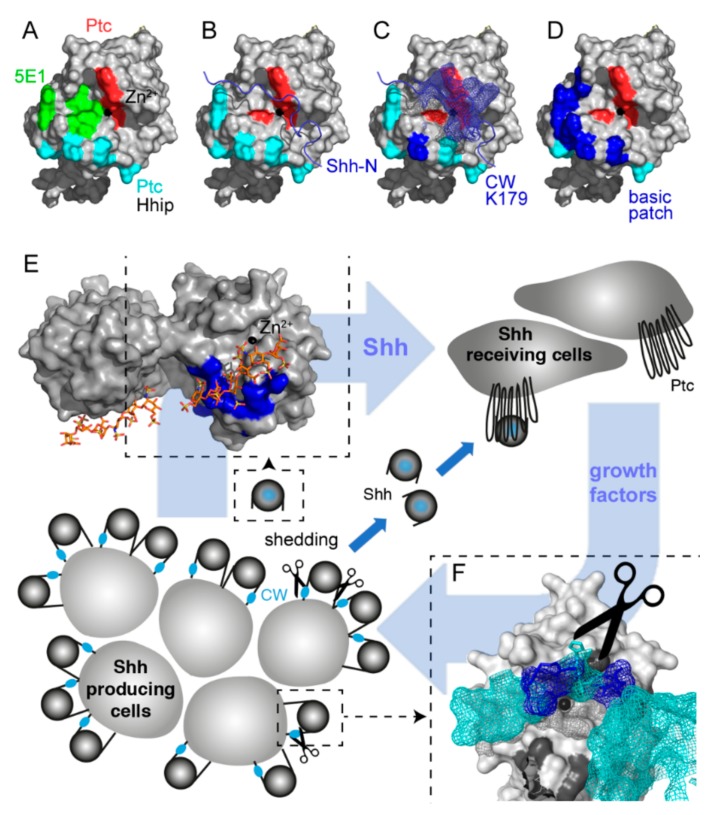
Sonic hedgehog (Shh) signaling plays a tumor-promoting role in many epithelial cancers. Cancer cells produce soluble a Shh that signals to distant stromal cells that express the receptor Patched (Ptc). These receiving cells respond by producing other soluble factors that promote cancer cell growth, generating a positive feedback loop. To interfere with reinforced Shh signaling, we examined the potential of defined heparin and heparan sulfate (HS) polysaccharides to block Shh solubilization and Ptc receptor binding. We confirm in vitro and in vivo that proteolytic cleavage of the N-terminal Cardin-Weintraub (CW) amino acid motif is a prerequisite for Shh solubilization and function. Consistent with the established binding of soluble heparin or HS to the Shh CW target motif, both polysaccharides impaired proteolytic Shh processing and release from source cells. We also show that HS and heparin bind to, and block, another set of basic amino acids required for unimpaired Shh binding to Ptc receptors on receiving cells. Both modes of Shh activity downregulation depend more on HS size and overall charge than on specific HS sulfation modifications. We conclude that heparin oligosaccharide interference in the physiological roles of HS in Shh release and reception may be used to expand the field of investigation to pharmaceutical intervention of tumor-promoting Shh functions.

Keywords: hedgehog; heparan sulfate; heparin.


Conflict of interest statement

The authors declare no conflicts of interest.

Figures

Figure 1
Figure 1
Multiple molecular interactions converge at the sonic hedgehog (Shh) pseudoactive site. (A) Shh contains a tetrahedrally coordinated Zn2+ cation (black sphere) that, together with amino acids H133, H134, H140, H180, and H182 (red), directly interacts with Hh-interacting protein (Hhip) and Patched (Ptc) peptides [52,53,54]. Cyan: amino acids K46, N51, R154, R156, and K179 that when mutated also affect Ptc binding [56]. The monoclonal antibody 5E1 binds to amino acids overlapping with the pseudoactive site (green) [55]. (B) In Hh clusters at the cell surface, unprocessed N-terminal peptides block the Shh binding site for Ptc (blue ribbon) [15,17]. (C) Unprocessed Shh clusters at the cell surface interact with heparan sulfate (HS) chains at two main sites: the basic N-terminal Cardin–Weintraub (CW) motif (mesh representation) and K179 (blue). (D) In addition to K179, processed bioactive Shh shows highly conserved basic amino acids in its vicinity (K88, R124, R154, R156, and K179, blue patch). (E) Model of two-way communication between tumor cells and their microenvironment (blue arrows). Tumors can secrete Shh that binds to Ptc receptors on stromal cells [35]. Stromal cells, including blood vessel cells, epithelial cells, fibroblasts, and immune cells, in turn support tumor growth by the secretion of other growth-promoting proteins [35,57]. Modified from [58]. Top left: clusters of basic amino acids at the molecular surface of soluble Shh can interact with HS (labeled blue are K88, R124, R154, R156, and K179; stick representation shows associated heparin in the structure). Analogous to Shh activity inhibition by MoAb 5E1, heparin/HS bound to this site may impair Shh binding to Ptc. (F) CW residues K32, R33, R34, K37, and K38 located in the N-terminal peptide of surface-associated Hh multimers (blue mesh) can also bind to HS. This would impair proteolytic processing of this site and Shh signaling to the tumor stroma. Shh: Sonic hedgehog, Ptc: Patched, Hhip: Hh-interacting protein, CW: Cardin–Weintraub motif.
Figure 2
Figure 2
Proteolytic processing of N-terminal CW residues is a prerequisite for controlled Shh and Drosophila Hh release and biofunction. (A) Targeted deletion of the CW motif (ShhΔ) diminishes protein release from the cell surface, but a protein with all five basic CW amino acids (blue) replaced by alanines (red, Shh5xA) is strongly released even in the absence of Scube2. Shh5xA + Scube2 was set to 100% and all other values are expressed relative to this value. One-way ANOVA; shown are average values ± SD, n = 4 for each data set. (B) UAS-CD8-GFP (green) produced in the anterior compartment or outside of the wing pouch under the control of GMR45433 and GMR45105, or in the posterior compartment under GMR48462 control. Cells in the posterior compartment produce endogenous Hh (red). Scale bar: 100 μm. (C) Adult Drosophila wings. In the normal wild-type situation (>CD8-GFP), the wing blade shows five longitudinal veins, an anterior cross vein, and a posterior cross vein. GMR-Gal4-induced Hh and Hh3xA expression (note the presence of only three basic arginines in the fly CW-motif, labeled blue) causes anterior wing overgrowth or leads to overgrowth of the wing costa (arrows). In contrast, GMR-Gal4 overexpression of HhΔ did not cause such defects, confirming biological inactivity of the expressed protein. Scale bar: 1 mm.
Figure 3
Figure 3
Heparin-modulated Shh release. (A) Shh expressed in Panc1 cells is bioactive, as indicated by Hh-dependent C3H10T1/2 reporter cell differentiation into alkaline phosphatase-producing osteoblasts. Shh biofunction was specifically inhibited by 5E1 and cyclopamine. *** denotes statistical significance (p < 0.0001, n = 2). (B) Increasing amounts of soluble heparin reduced Shh processing from Panc1 cells in a dose-dependent manner. * and ** denote statistical significance (p < 0.05 and p < 0.005, respectively, n = 5–8). (C) Impaired Shh release was specifically due to a blockade of the CW processing site because the release of Shh5xA lacking all HS-binding basic amino acids remained unaffected. n.s.: not significant (p > 0.05, n = 5–10). Shh release from Bosc23 cells (D) and HeLa cells (E) was also inhibited by increasing heparin concentrations. (F) Small heparin oligosaccharides (dp12 and dp30) variably reduce Shh release from Panc1 cells. Bottom: heparin structure. Most acetyl groups from GlcNAc residues are replaced by sulfate groups to generate an extended N-sulfated (NS) domain. Extensive subsequent modifications, such as epimerization of GlcA to IdoA and 2-O, 6-O, and 3-O sulfations generate a highly sulfated, negatively charged region. Xyl: xylose, Gal: galactose, GlcA: glucuronic acid, GlcNAc: N-acetylglucosamine, IdoA: iduronic acid. Modified after [69,70].
Figure 4
Figure 4
Soluble heparin impairs Ptc binding and biofunction of Shh. (A) Increasing amounts of soluble heparin were added to supernatants of Shh-expressing Panc1 cells. This significantly impaired Shh induced C3H10T1/2 precursor cell differentiation. *** p < 0.001 in all cases compared with C3H10T1/2 differentiation in the absence of heparin, n = 6–12. (B) Purmorphamine-dependent C3H10T1/2 differentiation is not impaired by 2–20 μg/mL exogenous heparin. ** p < 0.01, * p < 0.05, n.s. p > 0.05, n = 3–9. (C) Charge-dependent inhibition of C3H10T1/2 precursor cell differentiation. HSI carries the lowest charge density and heparin the highest charge density. Size-dependent inhibition of C3H10T1/2 precursor cell differentiation. Compared with that of heterogeneous heparin and dp30 heparin, the inhibitory potential of dp12 heparin is reduced. *** p < 0.001, * p < 0.05, n = 4–16.
Figure 5
Figure 5
HS microarray analysis of Shh binding. A total of 53 HS oligosaccharides were printed on microarray chips (36 dots/oligosaccharide) and incubated with soluble Shh. Shh binding is expressed as a histogram of relative fluorescence intensity. The numbered oligosaccharide sequences and structures for each sample are listed in the Supplementary Materials section; numbers in italics denote the average degree of sulfation per monosaccharide. (A) When 6-mers were compared, Shh binding to highly sulfated samples #33 and #39 was increased over that of all other samples (p < 0.001, n = 36). (B) Shh binding to 7-mers was also sulfation-dependent: Shh binding to samples #3, #9, and #25 was significantly increased over all other forms (p < 0.001, n = 36). (C) Sulfation-dependent Shh binding to oligosaccharides composed of 9 monosaccharides. Note that overall sulfation, but not any specific type of sulfation, is required for strong Shh binding (p < 0.001 when #40 is compared with all other samples, n = 36). (D) Sulfation-dependent Shh binding to oligosaccharides composed of 12 monosaccharides. All oligosaccharides differ significantly from each other in their Shh binding capacities (p < 0.001, n = 36), with the exception of samples #18 and #21 (n.s., p > 0.05) and #17 and #21 (p < 0.01, n = 36). (E) If oligosaccharides of different lengths but similar overall degrees of sulfation are compared, Shh binding to 12-mers is significantly increased over that of shorter forms. Longer oligosaccharides with reduced sulfation are less effective in Shh binding (compare #19 with #26). (F) If oligosaccharides of the same lengths but different overall degrees of sulfation are compared, Shh binding to highly sulfated probe #21 is preferred (p < 0.001, n = 36). Statistical significance was calculated by one-way ANOVA and Bonferroni’s multiple comparison test.

Similar articles

Cited by

References

    1. Ahn S., Joyner A.L. Dynamic changes in the response of cells to positive hedgehog signaling during mouse limb patterning. Cell. 2004;118:505–516. doi: 10.1016/j.cell.2004.07.023. - DOI - PubMed
    1. Briscoe J., Chen Y., Jessell T.M., Struhl G. A hedgehog-insensitive form of patched provides evidence for direct long-range morphogen activity of sonic hedgehog in the neural tube. Mol. Cell. 2001;7:1279–1291. doi: 10.1016/S1097-2765(01)00271-4. - DOI - PubMed
    1. Charron F., Stein E., Jeong J., McMahon A.P., Tessier-Lavigne M. The morphogen sonic hedgehog is an axonal chemoattractant that collaborates with netrin-1 in midline axon guidance. Cell. 2003;113:11–23. doi: 10.1016/S0092-8674(03)00199-5. - DOI - PubMed
    1. Harfe B.D., Scherz P.J., Nissim S., Tian H., McMahon A.P., Tabin C.J. Evidence for an expansion-based temporal Shh gradient in specifying vertebrate digit identities. Cell. 2004;118:517–528. doi: 10.1016/j.cell.2004.07.024. - DOI - PubMed
    1. Peters C., Wolf A., Wagner M., Kuhlmann J., Waldmann H. The cholesterol membrane anchor of the Hedgehog protein confers stable membrane association to lipid-modified proteins. Proc. Natl. Acad. Sci. USA. 2004;101:8531–8536. doi: 10.1073/pnas.0308449101. - DOI - PMC - PubMed

MeSH terms

LinkOut - more resources