. 2019 Apr 23;11(4):907.
doi: 10.3390/nu11040907.

Delayed-Type Hypersensitivity Underlying Casein Allergy Is Suppressed by Extracellular Vesicles Carrying miRNA-150

Affiliations

Delayed-Type Hypersensitivity Underlying Casein Allergy Is Suppressed by Extracellular Vesicles Carrying miRNA-150

Magdalena Wąsik et al. Nutrients. .

Abstract

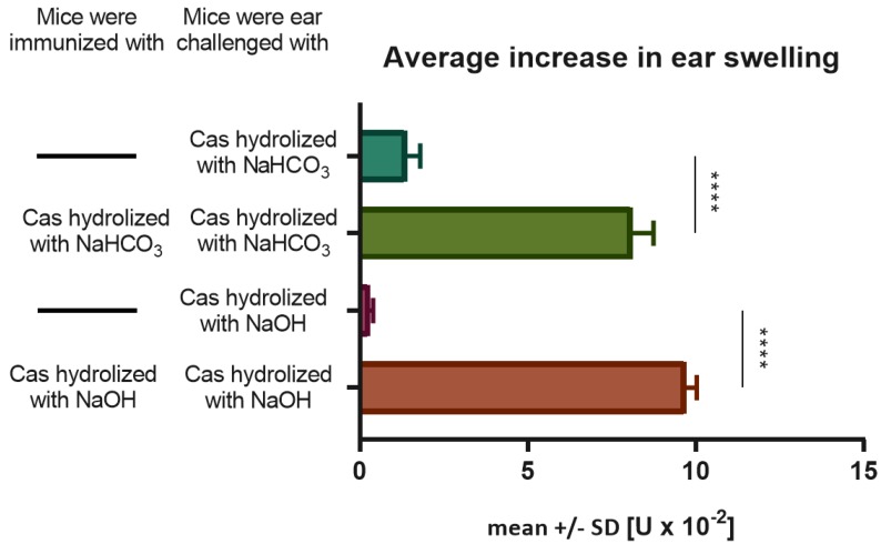
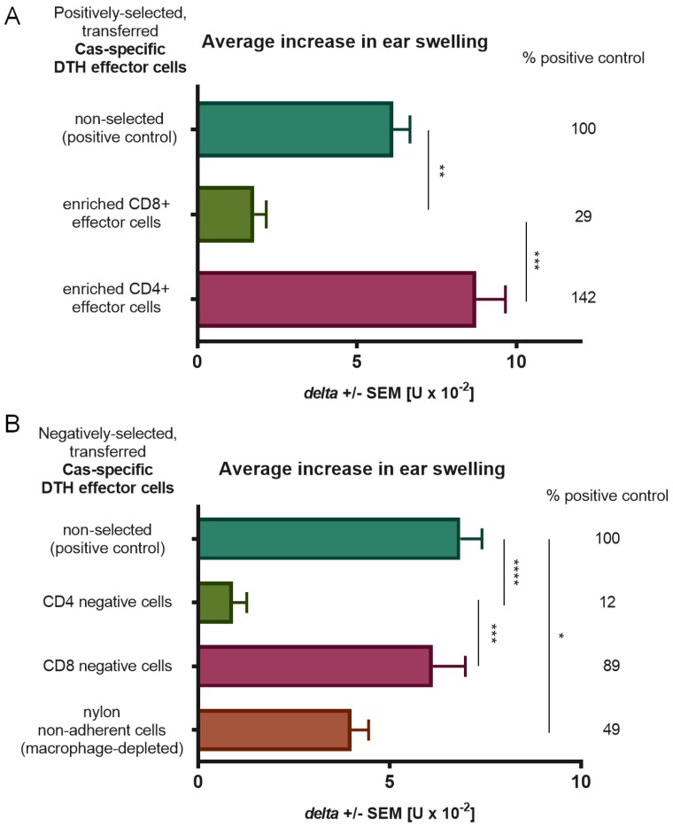
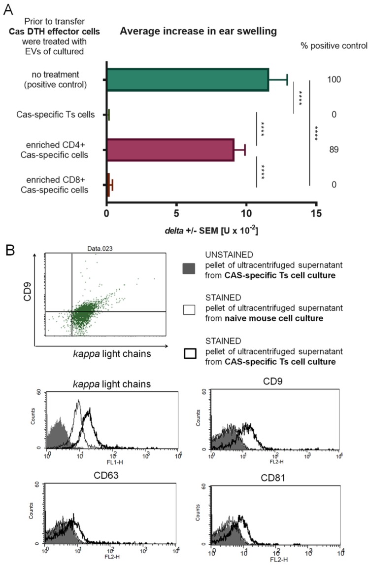
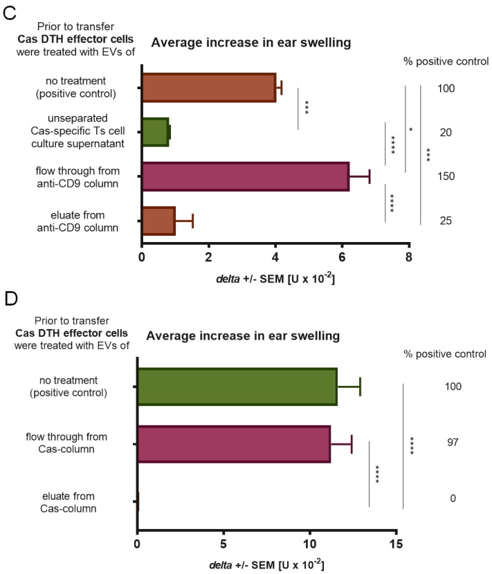
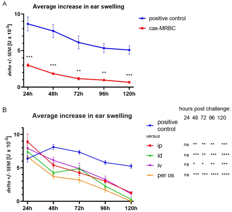
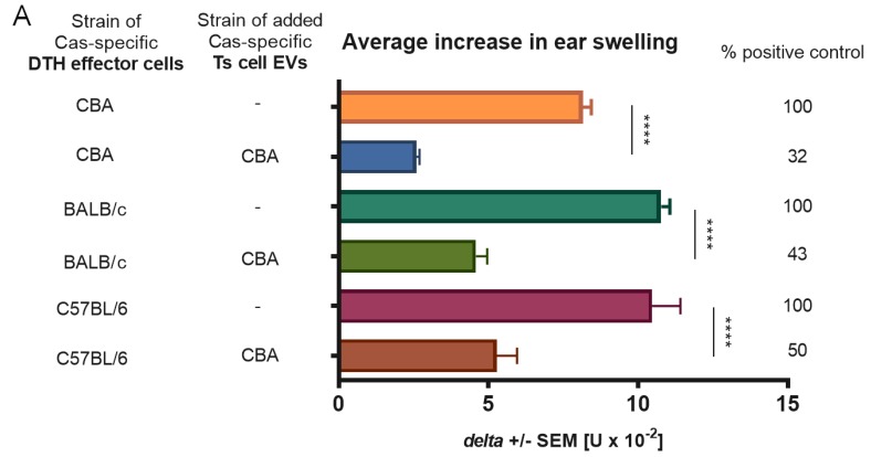
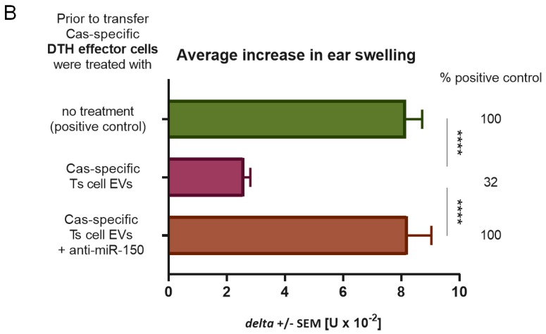
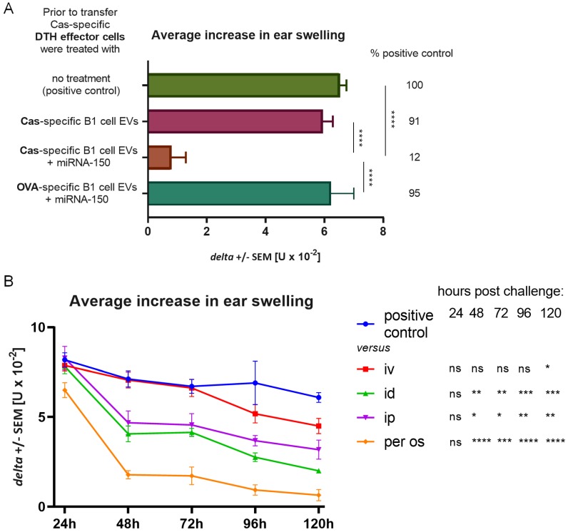
In patients with non-IgE-mediated milk allergy, a cellular mechanism of delayed-type hypersensitivity (DTH) is considered. Recent findings prove that cell-mediated reactions can be antigen-specifically inhibited by extracellular vesicles (EVs) carrying miRNA-150. We sought to establish a new mouse model of DTH to casein and test the possibility of antigen-specific suppression of the inflammatory reaction. To produce soluble antigenic peptides, casein was subjected to alkaline hydrolysis. DTH reaction to casein was induced in CBA, C57BL/6, and BALB/c mice by intradermal (id) injection of the antigen. Cells collected from spleens and lymph nodes were positively or negatively selected and transferred to naive recipients intravenously (iv). CBA mice were tolerized by iv injection of mouse erythrocytes conjugated with casein antigen and following id immunization with the same antigen. Suppressive EVs were harvested from cell cultures and serum of tolerized donors by means of ultrafiltration and ultracentrifugation for further therapeutic utilization. The newly established mouse model of DTH to casein was mediated by CD4+ Th1 cells and macrophages, while EVs produced by casein-tolerized animals effectively suppressed effector cell response, in an miRNA-150-dependent manner. Altogether, our observations contribute to the current understanding of non-IgE-mediated allergy to casein and of the possibilities to downregulate this reaction.

Keywords: casein; cell-mediated reactions; cow’s milk allergy; delayed-type hypersensitivity; extracellular vesicles; miRNA-150.


Conflict of interest statement

The authors declare no conflict of interest.

Figures

Figure 1
Figure 1
Immunogenicity of soluble casein (Cas) antigen obtained by alkaline hydrolysis with either NaHCO3 or NaOH. Mice had been intradermally (id) immunized with a saline solution of soluble Cas antigen (100 µg per mouse) 5 days before challenging by id administration of the same Cas solution (5 µg per earlobe). Twenty-four hours later ear swelling response was measured and expressed as mean ± SD [units (U) × 10−2 mm] (n = 4, N = 3). **** p < 0.0001.
Figure 2
Figure 2
Phenotyping of effector cells of delayed-type hypersensitivity (DTH) to soluble casein (Cas) antigen. Mice had been intradermally (id) immunized with a saline solution of soluble Cas antigen (100 µg per mouse) 5 days before harvest of lymph nodes and spleens containing effector cells, which were then subjected to positive (A) or negative (B) selection assays by, respectively, magnetic-advanced cell sorting or depletion with either monoclonal antibodies and complement or nylon wool separation. Afterwards, selected effector cells were transferred to naive recipients, which 24 h later were challenged by id administration of the same Cas solution (5 µg per earlobe). After 24 h, ear swelling response was measured and expressed as delta ± SEM [units (U) × 10−2 mm] (n = 5, N = 2), * p < 0.05; ** p < 0.01; *** p < 0.001; **** p < 0.0001.
Figure 3
Figure 3
Suppression of delayed-type hypersensitivity (DTH) to soluble casein (Cas) antigen is mediated by CD8+ suppressor T (Ts) cell-derived extracellular vesicles (EVs) expressing CD9 and CD81 tetraspanins and Cas-specific antibody light chains. (A) Mice had been intravenously administered with Cas-coupled syngeneic red blood cells, followed by intradermal (id) immunization with a saline solution of soluble Cas antigen (100 µg per mouse) 3 days before harvest of lymph nodes and spleens containing Ts cells, which were then subjected to positive selection assay, i.e., magnetic-advanced cell sorting, and cultured for subsequent 48 h. The resulting supernatant was filtered and ultracentrifuged to concentrate Ts cell EVs, used to treat DTH effector cells prior to adoptive transfer to recipients, challenged 24 h later by id administration of the same Cas solution (5 µg per earlobe). After 24 h, ear swelling response was measured and expressed as delta ± SEM [units (U) × 10−2 mm] (n = 5, N = 2). (B) Ts cell EVs were coated onto latex beads, stained with monoclonal antibodies against CD9, CD63, and CD81 tetraspanins or against mouse antibody kappa light chains, and then analyzed by flow cytometry (n = 3, N = 2). (C) Ts cell EVs were separated by antigen affinity chromatography on column filled with Sepharose coated with anti-CD9 monoclonal antibodies, and the resulting fractions, i.e., flow through or eluate, were used to treat DTH effector cells prior to adoptive transfer to recipients, challenged 24 h later by id administration of the same Cas solution (5 µg per earlobe). After 24 h, ear swelling response was measured and expressed as delta ± SEM [units (U) × 10−2 mm] (n = 5, N = 2). (D) Ts cell EVs were separated by antigen affinity chromatography on column filled with Cas-coated Sepharose, and the resulting fractions, i.e., flow through or eluate, were used to treat DTH effector cells prior to adoptive transfer to recipients, challenged 24 h later by id administration of the same Cas solution (5 µg per earlobe). After 24 h, ear swelling response was measured and expressed as delta ± SEM [units (U) × 10−2 mm] (n = 5, N = 3). * p < 0.05; ** p < 0.01; *** p < 0.001; **** p < 0.0001.
Figure 3
Figure 3
Suppression of delayed-type hypersensitivity (DTH) to soluble casein (Cas) antigen is mediated by CD8+ suppressor T (Ts) cell-derived extracellular vesicles (EVs) expressing CD9 and CD81 tetraspanins and Cas-specific antibody light chains. (A) Mice had been intravenously administered with Cas-coupled syngeneic red blood cells, followed by intradermal (id) immunization with a saline solution of soluble Cas antigen (100 µg per mouse) 3 days before harvest of lymph nodes and spleens containing Ts cells, which were then subjected to positive selection assay, i.e., magnetic-advanced cell sorting, and cultured for subsequent 48 h. The resulting supernatant was filtered and ultracentrifuged to concentrate Ts cell EVs, used to treat DTH effector cells prior to adoptive transfer to recipients, challenged 24 h later by id administration of the same Cas solution (5 µg per earlobe). After 24 h, ear swelling response was measured and expressed as delta ± SEM [units (U) × 10−2 mm] (n = 5, N = 2). (B) Ts cell EVs were coated onto latex beads, stained with monoclonal antibodies against CD9, CD63, and CD81 tetraspanins or against mouse antibody kappa light chains, and then analyzed by flow cytometry (n = 3, N = 2). (C) Ts cell EVs were separated by antigen affinity chromatography on column filled with Sepharose coated with anti-CD9 monoclonal antibodies, and the resulting fractions, i.e., flow through or eluate, were used to treat DTH effector cells prior to adoptive transfer to recipients, challenged 24 h later by id administration of the same Cas solution (5 µg per earlobe). After 24 h, ear swelling response was measured and expressed as delta ± SEM [units (U) × 10−2 mm] (n = 5, N = 2). (D) Ts cell EVs were separated by antigen affinity chromatography on column filled with Cas-coated Sepharose, and the resulting fractions, i.e., flow through or eluate, were used to treat DTH effector cells prior to adoptive transfer to recipients, challenged 24 h later by id administration of the same Cas solution (5 µg per earlobe). After 24 h, ear swelling response was measured and expressed as delta ± SEM [units (U) × 10−2 mm] (n = 5, N = 3). * p < 0.05; ** p < 0.01; *** p < 0.001; **** p < 0.0001.
Figure 4
Figure 4
Suppression of delayed-type hypersensitivity (DTH) to soluble casein (Cas) antigen is induced by intravenous (iv) injection of Cas-coupled syngeneic red blood cells and by CD8+ suppressor T (Ts) cell-derived extracellular vesicles (EVs) administered via different routes. (A) Mice had been administered iv with Cas-coupled syngeneic red blood cells, which was followed by intradermal (id) immunization with a saline solution of soluble Cas antigen (100 µg per mouse) 5 days before challenging by id administration of the same Cas solution (5 µg per earlobe). Subsequent ear swelling response was measured daily up to 120 h after challenge and expressed as delta ± SEM [units (U) × 10−2 mm] (n = 5, N = 2). (B) Mice had been id immunized with a saline solution of soluble Cas antigen (100 µg per mouse) 5 days before challenging by id administration of the same Cas solution (5 µg per earlobe). After measurement of 24 h ear swelling, Ts cell EVs were administered intraperitoneally (ip), id, iv, or per os, and subsequent ear swelling response was measured daily up to 120 h after challenge and expressed as delta ± SEM [units (U) × 10−2 mm] (n = 5, N = 2). * p < 0.05; ** p < 0.01; *** p < 0.001; **** p < 0.0001.
Figure 5
Figure 5
Major histocompatibility complex (MHC) restriction and dependence on miRNA-150 of suppression of delayed-type hypersensitivity (DTH) to soluble casein (Cas) antigen. (A) Mice of CBA, BALB/c, or C57BL/6 strains had been intradermally (id) immunized with a saline solution of soluble Cas antigen (100 µg per mouse) 5 days before harvest of lymph nodes and spleens containing effector cells, which were then treated with Cas-specific suppressor T (Ts) cell-derived extracellular vesicles (EVs) of tolerized CBA mice. Afterwards, effector cells were transferred to naive recipients of respective strain, which 24 h later were challenged by id administration of the same Cas solution (5 µg per earlobe). After 24 h, ear swelling response was measured and expressed as delta ± SEM [units (U) × 10−2 mm] (n = 4, N = 2). (B) Part of Ts cell EVs was incubated with miRNA-150 antagonist, i.e., anti-miR-150, prior to treatment of DTH effector cells, which were then adoptively transferred to recipients, challenged 24 h later by id administration of the same Cas solution (5 µg per earlobe). After 24 h, ear swelling response was measured and expressed as delta ± SEM [units (U) × 10−2 mm] (n = 5, N = 3). **** p < 0.0001.
Figure 5
Figure 5
Major histocompatibility complex (MHC) restriction and dependence on miRNA-150 of suppression of delayed-type hypersensitivity (DTH) to soluble casein (Cas) antigen. (A) Mice of CBA, BALB/c, or C57BL/6 strains had been intradermally (id) immunized with a saline solution of soluble Cas antigen (100 µg per mouse) 5 days before harvest of lymph nodes and spleens containing effector cells, which were then treated with Cas-specific suppressor T (Ts) cell-derived extracellular vesicles (EVs) of tolerized CBA mice. Afterwards, effector cells were transferred to naive recipients of respective strain, which 24 h later were challenged by id administration of the same Cas solution (5 µg per earlobe). After 24 h, ear swelling response was measured and expressed as delta ± SEM [units (U) × 10−2 mm] (n = 4, N = 2). (B) Part of Ts cell EVs was incubated with miRNA-150 antagonist, i.e., anti-miR-150, prior to treatment of DTH effector cells, which were then adoptively transferred to recipients, challenged 24 h later by id administration of the same Cas solution (5 µg per earlobe). After 24 h, ear swelling response was measured and expressed as delta ± SEM [units (U) × 10−2 mm] (n = 5, N = 3). **** p < 0.0001.
Figure 6
Figure 6
Suppression of delayed-type hypersensitivity (DTH) to soluble casein (Cas) antigen by miRNA-150-supplemented B1 cell-derived extracellular vesicles (EVs) of Cas-immunized mice. (A) Mice had been intradermally (id) immunized with a saline solution of soluble Cas antigen or ovalbumin (OVA, both 100 µg per mouse) 3 days before harvest of lymph nodes and spleens containing B1 cells, which were cultured for a subsequent 48 h. The resulting supernatant was filtered and ultracentrifuged to concentrate Cas-specific or OVA-specific B1 cell EVs, then supplemented with miRNA-150 prior to treatment of Cas-specific DTH effector cells, which were adoptively transferred to recipients, challenged 24 h later by id administration of the same Cas solution (5 µg per earlobe). After 24 h, ear swelling response was measured and expressed as delta ± SEM [units (U) × 10−2 mm] (n = 5, N = 2). (B) Mice had been id immunized with a saline solution of soluble Cas antigen (100 µg per mouse) 5 days before challenging by id administration of the same Cas solution (5 µg per earlobe). After measurement of 24 h ear swelling, miRNA-150-supplemented, Cas-specific B1 cell EVs were administered intraperitoneally (ip), id, intravenously (iv), or per os, and subsequent ear swelling response was measured daily up to 120 h after challenge and expressed as delta ± SEM [units (U) × 10−2 mm] (n = 4, N = 2). * p < 0.05; ** p < 0.01; *** p < 0.001; **** p < 0.0001.

Similar articles

Cited by

References

    1. Wąsik M., Nazimek K., Bryniarski K. Allergic reactions to cow’s milk: Pathomechanism, diagnostic and therapeutic strategies, possibilities of food tolerance induction. Postepy Hig. Med. Dosw. 2018;72:1–11. doi: 10.5604/01.3001.0011.8254. (In Polish) - DOI
    1. Hill D.J., Ball G., Hosking C.S. Clinical manifestations of cows’ milk allergy in childhood. I. Associations with in-vitro cellular immune responses. Clin. Allergy. 1988;18:469–479. doi: 10.1111/j.1365-2222.1988.tb02897.x. - DOI - PubMed
    1. Hill D., Firer M., Shelton M., Hosking C. Manifestations of milk allergy in infancy: Clinical and immunologic findings. J. Pediatr. 1986;109:270–276. doi: 10.1016/S0022-3476(86)80384-5. - DOI - PubMed
    1. Díaz M., Guadamuro L., Espinosa-Martos I., Mancabelli L., Jiménez S., Molinos-Norniella C., Pérez-Solis D., Milani C., Rodríguez J.M., Ventura M., et al. Microbiota and Derived Parameters in Fecal Samples of Infants with Non-IgE Cow’s Milk Protein Allergy under a Restricted Diet. Nutrients. 2018;10:1481. doi: 10.3390/nu10101481. - DOI - PMC - PubMed
    1. Nowak-Węgrzyn A., Katz Y., Mehr S.S., Koletzko S. Non-IgE-mediated gastrointestinal food allergy. J. Allergy Clin. Immunol. 2015;135:1114–1124. doi: 10.1016/j.jaci.2015.03.025. - DOI - PubMed