. 2019 Jul;32(4):211-220.
doi: 10.1177/0891988719841728. Epub 2019 Apr 24.

Analysis of Surface Levels of IL-1 Receptors and Macrophage Scavenger Receptor I in Peripheral Immune Cells of Patients With Alzheimer Disease

Affiliations

Analysis of Surface Levels of IL-1 Receptors and Macrophage Scavenger Receptor I in Peripheral Immune Cells of Patients With Alzheimer Disease

Philipp Spitzer et al. J Geriatr Psychiatry Neurol. 2019 Jul.

Abstract

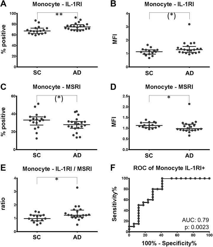
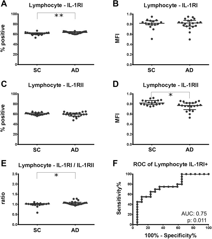
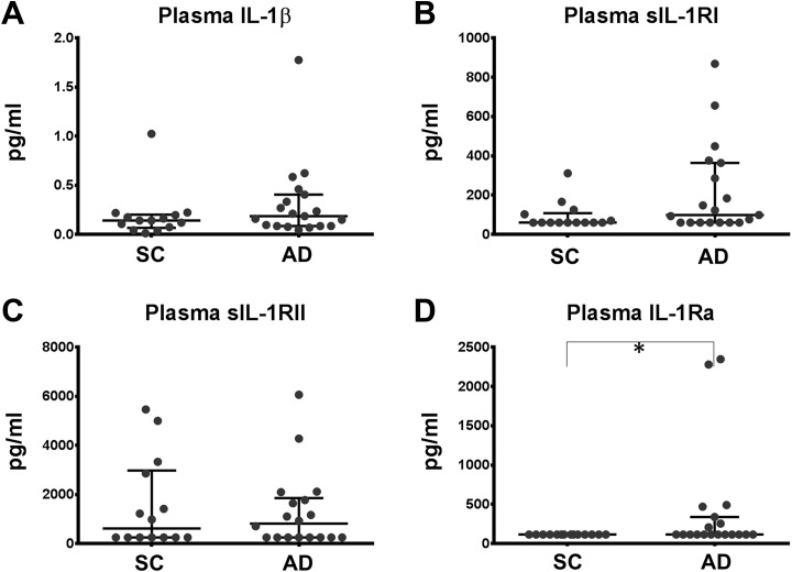
Increased concentrations of interleukin 1 (IL-1) in the cerebrospinal fluid and serum of patients with Alzheimer disease (AD) reduced phagocytic capacity point to an inflammatory activation of mononuclear phagocytes in AD. Interleukin 1 receptors (IL-1R) and the macrophage scavenger receptor I (MSRI) are important players in IL-1 signaling and phagocytosis. In 20 patients with AD and 17 controls, IL-1RI, IL-1RII, and MSRI were assessed on peripheral blood mononuclear cells by flow cytometry. IL-1β, soluble IL-1 receptors, and IL-1R antagonist (IL-1Ra) were measured by enzyme-linked immunosorbent assay. The fraction of IL-1RI+ monocytes was increased by 10% and the expression of MSRI was reduced by 12% in AD. A 3.6% increased fraction of IL-1RI+ lymphocytes was accompanied by a 6.1% reduced expression of IL-1RII. The IL-1RI on monocytes and lymphocytes discriminated patients with AD with an accuracy of 0.79 and 0.75, respectively. The IL-1Ra was elevated in AD. Changes in the expression of IL-1 receptors and MSRI on peripheral blood cells fit to the concept of a proinflammatory state of the peripheral immune system. However, the observed differences are not strong enough to suggest their application as biomarkers for AD.

Keywords: Alzheimer disease; IL-1RI; MSRI; biomarker; dementia; neurodegeneration; neuroinflammation.


Conflict of interest statement

Declaration of Conflicting Interests: The author(s) declared the following potential conflicts of interest with respect to the research, authorship, and/or publication of this article: PL received consultation and lectures honoraria from Innogenetics, IBL International, and AJ Roboscreen.

Figures

Figure 1.
Figure 1.
Expression of IL-1RI is increased; expression of MSRI is decreased on monocytes of patients with AD. PBMC of patients with AD and SC was labeled with antibodies directed against IL-1RI and MSRI. A and C, Fraction of monocytes staining positive for IL-1RI and MSRI. B and D, The mean fluorescence intensity (MFI) of the IL-1RI and the MSRI on monocytes are depicted. The ratio of IL-1RI (MFI)/MSRI (MFI) comparing the 2 populations is plotted in (E). F, The receiver operating characteristic curve (ROC) for the discrimination of patients with and without AD by the fraction of IL-1RI-positive monocytes. Trend: *P < .10; significance: *P < .05; **P < .01. AD indicates Alzheimer disease; IL-1RI, interleukin 1 receptor subtype I; MSRI, macrophage scavenger receptor I; PBMC, peripheral blood mononuclear cell; SC, symptomatic controls.
Figure 2.
Figure 2.
Regression of IL-1RI, MSRI, and the IL-1RI/MSRI ratio in monocytes as well as IL-1RI, IL-1RII, and the IL-1RI/IL-1RII ratio on lymphocytes with age. All parameters are plotted versus age and linear regression lines were calculated separately for patients with AD (filled circles and continuous line) and symptomatic controls (empty circles, broken line). For none of the parameters, slopes differed significantly. The probability that the elevations of the 2 regression lines are identical is indicated as P value in the lower right corner of the diagram. AD indicates Alzheimer disease; IL-1RI, interleukin 1 receptor subtype I; IL-1RII, interleukin 1 receptor subtype II; MSRI, macrophage scavenger receptor I.
Figure 3.
Figure 3.
Expression of IL-1RI is increased and expression of IL-RII is decreased on lymphocytes of patients with AD. PBMC of patients with AD and SC was labeled with antibodies directed against IL-1RI and IL-1RII. A, C, Fraction of lymphocytes staining positive for IL-1RI and IL1-RII. B, D, The mean fluorescence intensity (MFI) of IL-1RI and IL-1RII on lymphocytes. The ratio of IL-1RI/IL-1RII comparing the 2 populations is plotted in (E). F, The receiver operating characteristic curve (ROC) for the discrimination of patients with or without AD by the fraction of IL-1RI-positive lymphocytes. Trend: *P < .10; significance: *P < .05; **P < .01. AD indicates Alzheimer disease; IL-1RI, interleukin 1 receptor subtype I; IL-1RII indicates interleukin 1 receptor subtype II; IL-1RII, interleukin 1 receptor subtype II; MSRI, macrophage scavenger receptor I; PBMC, peripheral blood mononuclear cell; SC, symptomatic controls.
Figure 4.
Figure 4.
Concentrations of IL-1β, sIL-1RI, sIL-1RII, and IL-1Ra in plasma of patients with AD. Levels of IL-1β, sIL-1RI, sIL-1RII, and IL-1Ra were quantified by ELISA in plasma. Results are depicted as median [interquartile ranges]. Significance was calculated with the nonparametric Mann-Whitney U test. Values below the lower limit of quantification were set to the lower limit of quantification (IL-1RI: 60 pg/mL, IL-1RII: 250 pg/mL, IL-1Ra: 115 pg/mL). *P < .05. AD indicates Alzheimer disease; IL-1Ra, interleukin 1 receptor antagonist; IL-1RI, interleukin 1 receptor subtype I; IL-1RII indicates interleukin 1 receptor subtype II; IL-1RII, interleukin 1 receptor subtype II; IL-1Ra, interleukin 1 receptor antagonist; SC, symptomatic controls; sIL-1RI, soluble interleukin 1 receptor subtype I; sIL-1RII, soluble interleukin 1 receptor subtype II.

Similar articles

Cited by

References

    1. Heneka MT, Carson MJ, El Khoury J, et al. Neuroinflammation in Alzheimer’s disease. Lancet Neurol. 2015;14(4):388–405. - PMC - PubMed
    1. Heppner FL, Ransohoff RM, Becher B. Immune attack: the role of inflammation in Alzheimer disease. Nat Rev Neurosci. 2015;16(6):358–372. - PubMed
    1. Simard AR, Soulet D, Gowing G, Julien JP, Rivest S. Bone marrow-derived microglia play a critical role in restricting senile plaque formation in Alzheimer’s disease. Neuron. 2006;49(4):489–502. - PubMed
    1. Patel NS, Paris D, Mathura V, Quadros AN, Crawford FC, Mullan MJ. Inflammatory cytokine levels correlate with amyloid load in transgenic mouse models of Alzheimer’s disease. J Neuroinflamm. 2005;2(1):9. - PMC - PubMed
    1. Stewart CR, Stuart LM, Wilkinson K, et al. CD36 ligands promote sterile inflammation through assembly of a Toll-like receptor 4 and 6 heterodimer. Nat Immunol. 2010;11(2):155–161. - PMC - PubMed

Publication types

Substances