

Expression of TLR2, TLR3, TLR4, and TLR7 on pulmonary lymphocytes of Schistosoma japonicum-infected C57BL/6 mice
- PMID: 31018808
- PMCID: PMC6830883
- DOI: 10.1177/1753425919840424
Expression of TLR2, TLR3, TLR4, and TLR7 on pulmonary lymphocytes of Schistosoma japonicum-infected C57BL/6 mice
Abstract
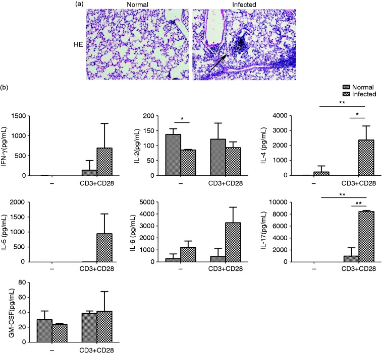
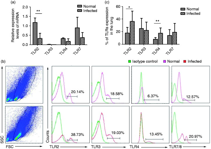
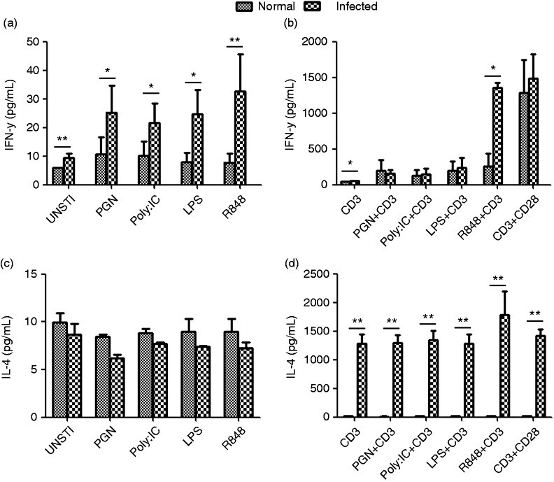
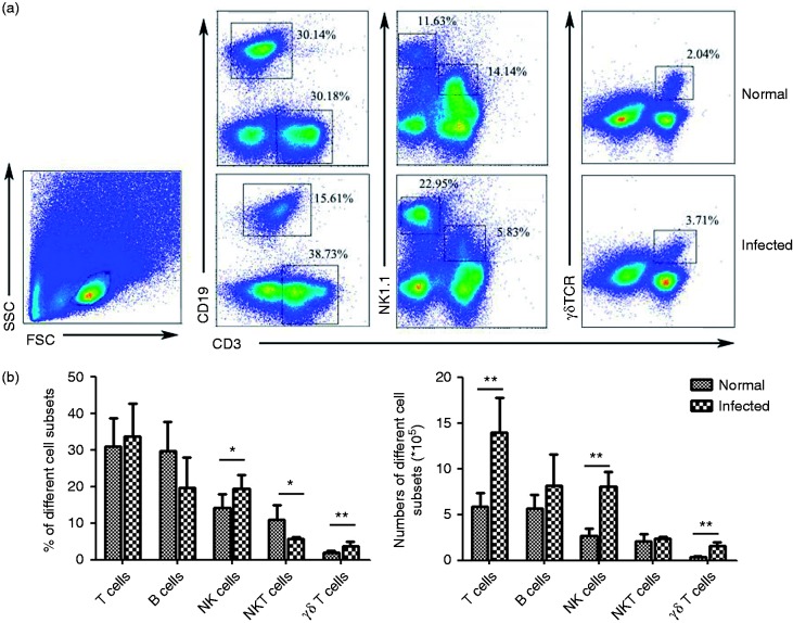
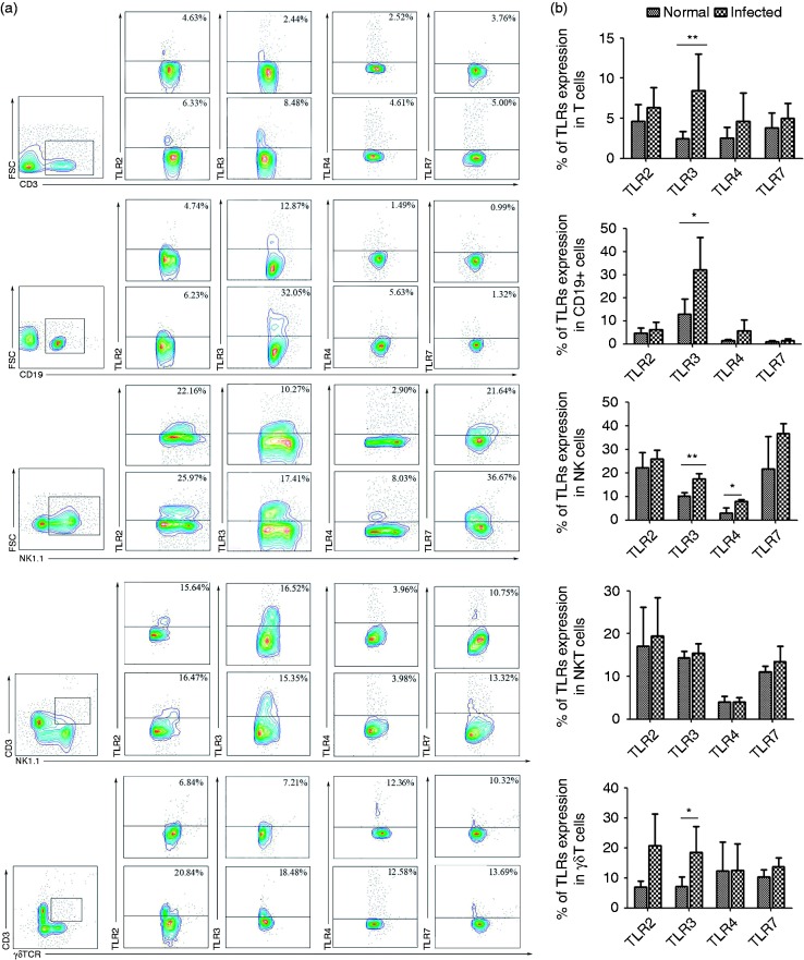
Despite the paramount role of TLRs in the induction of innate immune and inflammatory responses, there is a paucity of studies on the role of TLRs in Schistosoma japonicum infection. Here, we observed obvious infiltration of inflammatory cells in S. japonicum-infected C57BL/6 mouse lungs. Expression and release of IFN-γ, IL-4, and IL-17 were significantly higher in pulmonary lymphocytes from infected mice compared with control mice in response to anti-CD3 plus anti-CD28 mAbs. Higher percentages of TLR2, TLR3, TLR4, and TLR7 were expressed on such lymphocytes, and the TLR agonists PGN, Poly I:C, LPS, and R848 induced a higher level of IFN-γ. However, a higher level of IL-4 was found in the supernatant of pulmonary lymphocytes from infected mice stimulated by these TLR agonists plus CD3 Ab. Only R848 plus anti-CD3 mAb could induce a higher level of IFN-γ in such lymphocytes. TLR expressions were then compared on different pulmonary lymphocytes after infection, including T cells, B cells, NK cells, NKT cells, and γδT cells. The expression levels of TLR3 on T cells, B cells, NK cells, and γδT cells were increased in the lungs after infection. NK cells also expressed higher levels of TLR4 after infection of control mice. Collectively, these findings highlight the potential role of TLR expression in the context of S. japonicum infection.
Keywords: TLRs; cytokines; ligands; lung.
Conflict of interest statement
The author(s) declared no potential conflicts of interest with respect to the research, authorship, and/or publication of this article.
Figures
Similar articles
-
TLR7 Modulated T Cell Response in the Mesenteric Lymph Node of Schistosoma japonicum-Infected C57BL/6 Mice.J Immunol Res. 2019 Dec 22;2019:2691808. doi: 10.1155/2019/2691808. eCollection 2019. J Immunol Res. 2019. PMID: 31930147 Free PMC article.
-
TLR3 Modulates the Response of NK Cells against Schistosoma japonicum.J Immunol Res. 2018 Aug 30;2018:7519856. doi: 10.1155/2018/7519856. eCollection 2018. J Immunol Res. 2018. PMID: 30246036 Free PMC article.
-
miR-181a regulates the host immune response against Schistosoma japonicum infection through the TLR4 receptor pathway.Parasit Vectors. 2021 Oct 24;14(1):548. doi: 10.1186/s13071-021-05063-z. Parasit Vectors. 2021. PMID: 34689797 Free PMC article.
-
Type 1 cytokine/chemokine production by mouse NK cells following activation of their TLR/MyD88-mediated pathways.Int Immunol. 2007 Mar;19(3):311-20. doi: 10.1093/intimm/dxl148. Epub 2007 Feb 7. Int Immunol. 2007. PMID: 17289654
-
Roles of Th17 cells in pulmonary granulomas induced by Schistosoma japonicum in C57BL/6 mice.Cell Immunol. 2013 Sep-Oct;285(1-2):149-57. doi: 10.1016/j.cellimm.2013.09.008. Epub 2013 Oct 14. Cell Immunol. 2013. PMID: 24212062
Cited by
-
The Roles of Various Immune Cell Populations in Immune Response against Helminths.Int J Mol Sci. 2023 Dec 28;25(1):420. doi: 10.3390/ijms25010420. Int J Mol Sci. 2023. PMID: 38203591 Free PMC article. Review.
-
Evaluation of the TLR3 involvement during Schistosoma japonicum-induced pathology.BMC Immunol. 2024 Jan 3;25(1):2. doi: 10.1186/s12865-023-00586-9. BMC Immunol. 2024. PMID: 38172683 Free PMC article.
-
Expression profile of Toll-like receptors and cytokines in the cecal tonsil of chickens challenged with Eimeria tenella.Parasitol Res. 2024 Oct 10;123(10):347. doi: 10.1007/s00436-024-08371-2. Parasitol Res. 2024. PMID: 39387973
-
Expression of a stress-inducible heme oxygenase-1 in NK cells is maintained in the process of human aging.Front Immunol. 2024 Jul 19;15:1398468. doi: 10.3389/fimmu.2024.1398468. eCollection 2024. Front Immunol. 2024. PMID: 39100660 Free PMC article.
-
Changes of CD103-expressing pulmonary CD4+ and CD8+ T cells in S. japonicum infected C57BL/6 mice.BMC Infect Dis. 2019 Nov 27;19(1):999. doi: 10.1186/s12879-019-4633-8. BMC Infect Dis. 2019. PMID: 31775660 Free PMC article.
References
-
- Steinmann P, Keiser J, Bos R, et al. Schistosomiasis and water resources development: systematic review, meta-analysis, and estimates of people at risk. Lancet Infect Dis 2006; 6: 411–425. - PubMed
-
- Cha H, Qin W, Yang Q, et al. Differential pulmonic NK and NKT cell responses in Schistosoma japonicum-infected mice. Parasitol Res 2017; 116: 559–567. - PubMed
-
- Gause WC, Urban JJ, Stadecker MJ. The immune response to parasitic helminths: insights from murine models. Trends Immunol 2003; 24: 269–277. - PubMed
Publication types
MeSH terms
Substances
LinkOut - more resources
Full Text Sources
