Observational Study
. 2019 Apr 25;7(1):66.
doi: 10.1186/s40168-019-0679-5.

Chemotherapy-induced oral mucositis is associated with detrimental bacterial dysbiosis

Affiliations
Observational Study

Chemotherapy-induced oral mucositis is associated with detrimental bacterial dysbiosis

Bo-Young Hong et al. Microbiome. .

Abstract

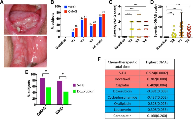
Background: Gastrointestinal mucosal injury (mucositis), commonly affecting the oral cavity, is a clinically significant yet incompletely understood complication of cancer chemotherapy. Although antineoplastic cytotoxicity constitutes the primary injury trigger, the interaction of oral microbial commensals with mucosal tissues could modify the response. It is not clear, however, whether chemotherapy and its associated treatments affect oral microbial communities disrupting the homeostatic balance between resident microorganisms and the adjacent mucosa and if such alterations are associated with mucositis. To gain knowledge on the pathophysiology of oral mucositis, 49 subjects receiving 5-fluorouracil (5-FU) or doxorubicin-based chemotherapy were evaluated longitudinally during one cycle, assessing clinical outcomes, bacterial and fungal oral microbiome changes, and epithelial transcriptome responses. As a control for microbiome stability, 30 non-cancer subjects were longitudinally assessed. Through complementary in vitro assays, we also evaluated the antibacterial potential of 5-FU on oral microorganisms and the interaction of commensals with oral epithelial tissues.

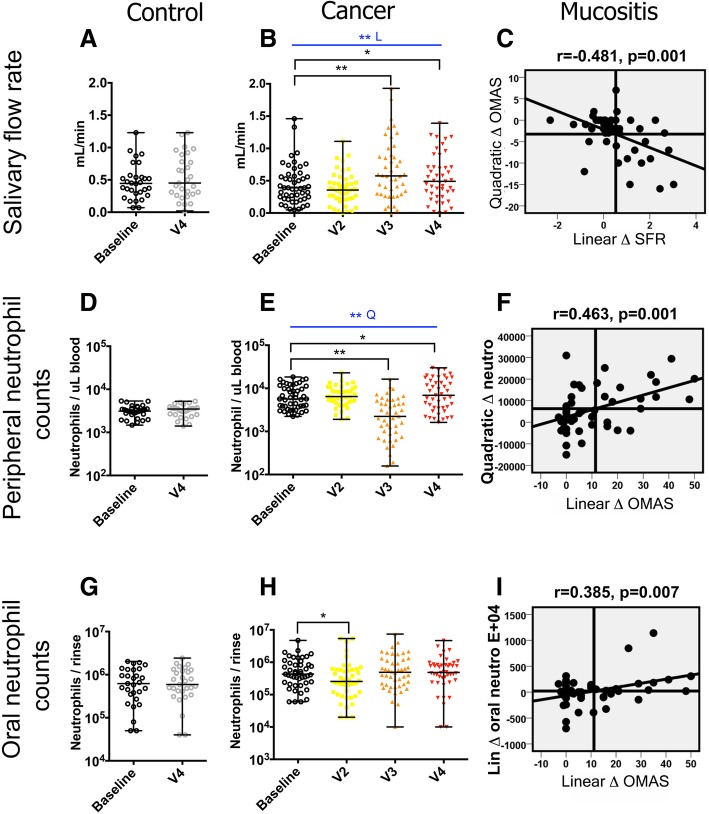
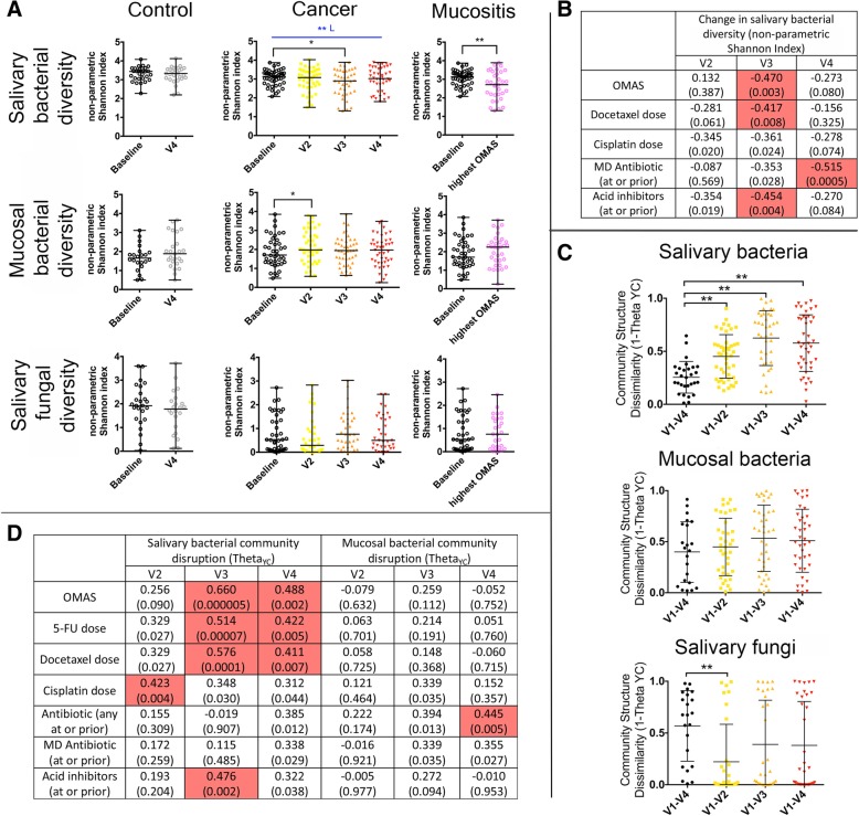
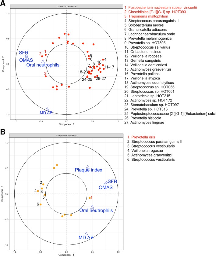
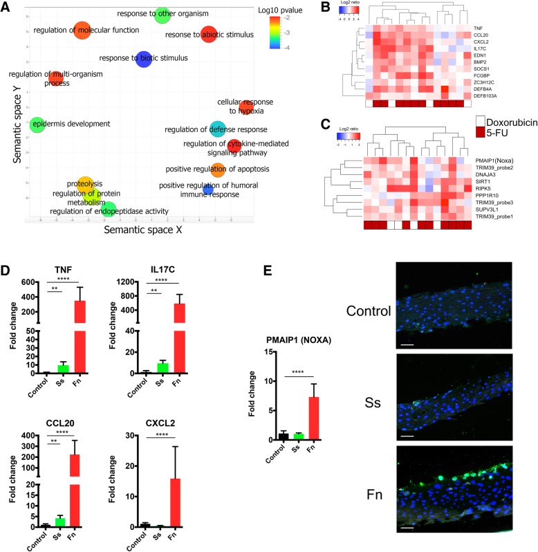
Results: Oral mucositis severity was associated with 5-FU, increased salivary flow, and higher oral granulocyte counts. The oral bacteriome was disrupted during chemotherapy and while antibiotic and acid inhibitor intake contributed to these changes, bacteriome disruptions were also correlated with antineoplastics and independently and strongly associated with oral mucositis severity. Mucositis-associated bacteriome shifts included depletion of common health-associated commensals from the genera Streptococcus, Actinomyces, Gemella, Granulicatella, and Veillonella and enrichment of Gram-negative bacteria such as Fusobacterium nucleatum and Prevotella oris. Shifts could not be explained by a direct antibacterial effect of 5-FU, but rather resembled the inflammation-associated dysbiotic shifts seen in other oral conditions. Epithelial transcriptional responses during chemotherapy included upregulation of genes involved in innate immunity and apoptosis. Using a multilayer epithelial construct, we show mucositis-associated dysbiotic shifts may contribute to aggravate mucosal damage since the mucositis-depleted Streptococcus salivarius was tolerated as a commensal, while the mucositis-enriched F. nucleatum displayed pro-inflammatory and pro-apoptotic capacity.

Conclusions: Altogether, our work reveals that chemotherapy-induced oral mucositis is associated with bacterial dysbiosis and demonstrates the potential for dysbiotic shifts to aggravate antineoplastic-induced epithelial injury. These findings suggest that control of oral bacterial dysbiosis could represent a novel preventive approach to ameliorate oral mucositis.

Keywords: Cancer; Chemotherapy; Microbiome; Mucosal-microbial crosstalk; Oral mucositis.


Conflict of interest statement

Ethics approval and consent to participate

This study was approved by the Institutional Review Board at UConn Health (IRB number IE-11-037 J-2). Written informed consent was received from all participants.

Consent for publication

Not applicable

Competing interests

The authors declare that they have no competing interests.

Publisher’s Note

Springer Nature remains neutral with regard to jurisdictional claims in published maps and institutional affiliations.

Figures

Fig. 1
Fig. 1
Incidence and clinical presentation of oral mucositis during chemotherapy and correlation with antineoplastics. a Intraoral images of a patient affected by oral mucositis. b Mucositis incidence according to the WHO scale, which evaluates in a categorical scale of 0 to 4 objective signs and patient-reported symptoms, and to the OMAS scale, which is based solely on objective signs of erythema and ulceration. OMAS scores reported here could range from 0 to 45 and represent the aggregated scores from nine intra-oral sites evaluated. c, d The clinical progression of mucositis in mucositis-positive subjects (n = 32 for the WHO scale and n = 37 for OMAS). Graphs show individual data points with median and range. ** indicates a p value < 0.01 and *** indicates a p value < 0.001 when comparing each time point to baseline via Wilcoxon matched-pairs signed rank tests. e Incidence of mucositis in subjects taking 5-FU and those on doxorubicin. * indicates a p value of < 0.05 when comparing incidence via chi-square. f Correlations between chemotherapeutic total drug doses and mucositis severity. Data represent Spearman correlation coefficients with p values in parenthesis. Colored cells show correlations significant after adjustment for multiple comparisons via the FDR method. Only drugs given to at least 15% of subjects were included in the analysis
Fig. 2
Fig. 2
Changes in salivary flow rate and peripheral and oral neutrophils during chemotherapy and correlation of these changes with oral mucositis severity. a, b Salivary flow rate (SFR) in control and cancer subjects. A statistically significant increase in SFR was seen in cancer subjects at V3 and V4 compared to baseline. Also, the linear change (L) in SFR during chemotherapy was significant. c A correlation between the negative quadratic change in OMAS (low, high, low) and the positive linear change in SFR in cancer subjects indicating SFR increased concomitant to or following mucositis. Each data point in the plot represents the change in a subject and was generated by transforming data from each visit according to orthogonal polynomial contrast coefficients followed by aggregation of the data from the four visits. d, e The change in peripheral neutrophils in control and cancer subjects. A statistically significant decrease during chemotherapy was seen at V3 and V4 compared to baseline. Also, the change during chemotherapy modelled with a quadratic polynomial contrast (Q) was significant. f A correlation between the positive quadratic change in peripheral neutrophils (high, low, high) and the positive linear change in OMAS indicating a correlation between neutrophil depletion and mucositis severity. g, h The change in oral neutrophils in control and cancer subjects. A statistically significant decrease was seen at V2 during chemotherapy. i A positive correlation between the linear change in oral neutrophils and mucositis severity. * indicates a p value < 0.05 and ** < 0.01
Fig. 3
Fig. 3
Changes in the oral microbiome during chemotherapy and in relation to the development of oral mucositis. a Bacterial and fungal microbiome diversity in control and cancer subjects. Salivary bacterial diversity decreased at visit 3 compared to baseline and also the linear change (L) in diversity was significant. Decreased salivary bacterial diversity was also seen at the visit with the highest OMAS. Mucosal bacterial diversity increased at V2, while no changes were seen in salivary fungal communities. b Variables significantly correlated with the change in salivary bacterial diversity at each visit when compared to baseline. Data represent Spearman correlation coefficients with p values in parenthesis. Colored cells show correlations significant after adjustment for multiple comparisons via the FDR method. c Changes in community structure as measured by the ThetaYC distance from baseline (V1) to each visit. Black data points indicate changes in control subjects and color data points indicate cancer subjects. ** indicates a p value < 0.01 when comparing to control subjects via Mann-Whitney Rank tests. d Variables significantly correlated with changes in salivary and mucosal bacterial community structure (1-ThetaYC distance from baseline to each visit). Data represent Spearman correlation coefficients with p values in parenthesis. Colored cells show correlations significant after adjustment for multiple comparisons via the FDR method
Fig. 4
Fig. 4
Longitudinal covariation of bacterial relative abundances and clinical signs of oral mucositis during chemotherapy. a A circle correlation plot depicting covariation of salivary bacterial abundances with oral mucositis severity (OMAS) and other significant clinical variables, as determined via multi-level sPLS analysis. Bacterial species appear as red circles and clinical variables are shown as blue triangles. Data points were placed in the plot according to their correlation with the two main components. Positively correlated variables follow the same direction from the origin. The greater the distance from the origin, the stronger the association. Only variables with a correlation coefficient greater than 0.4 are shown. Red numbers and corresponding names indicate bacterial species positively correlated with OMAS (enriched as mucositis severity increased), while black numbers and names indicate bacterial species negatively correlated with OMAS (depleted during severe mucositis). SFR salivary flow rate, MD AB multi-dose antibiotic intake; b a similar analysis done for mucosal bacterial taxa. Bacterial species appear as orange circles and clinical variables as blue triangles. Red numbers and names indicate species positively correlated with OMAS, while black numbers and names indicate bacterial species negatively correlated with OMAS
Fig. 5.
Fig. 5.
Epithelial responses to chemotherapeutic treatment and potential for commensals to modulate mucosal response. Changes in the oral epithelial transcriptome during chemotherapy (baseline to V3) were evaluated in 14 subjects via DASL-whole genome arrays. a A scatter plot showing significantly upregulated gene ontology (GO) terms summarized using REVIGO. Size of each circle represents GO term frequency (log scale) and color its p value (log scale), with lower p values in blue. b genes related to the immune response that were upregulated more than twofold during chemotherapy. c Genes related to apoptosis upregulated more than twofold during chemotherapy. d The expression of selected immune genes as evaluated via real-time PCR after exposure of a 3D multilayer oral epithelial construct to Streptococcus salivarius ATCC 9222 (Ss) or Fusobacterium nucleatum subsp. vincentii ATCC 49256 (Fn). e Expression of the proapoptotic gene PMAIP1 (NOXA) as measured via real-time PCR and micrographs depicting multilayer oral epithelial constructs stained with a fluorescent TUNEL assay to evaluate cell death. TUNEL-positive cells appear green and nuclei in blue. Bar = 50 μM

Similar articles

Cited by

References

    1. Vera-Llonch M, Oster G, Ford CM, Lu J, Sonis S. Oral mucositis and outcomes of allogeneic hematopoietic stem-cell transplantation in patients with hematologic malignancies. Support Care Cancer. 2007;15(5):491–496. doi: 10.1007/s00520-006-0176-9. - DOI - PubMed
    1. Keefe DM, Schubert MM, Elting LS, Sonis ST, Epstein JB, Raber-Durlacher JE, et al. Updated clinical practice guidelines for the prevention and treatment of mucositis. Cancer. 2007;109(5):820–831. doi: 10.1002/cncr.22484. - DOI - PubMed
    1. Murphy BA, Beaumont JL, Isitt J, Garden AS, Gwede CK, Trotti AM, et al. Mucositis-related morbidity and resource utilization in head and neck cancer patients receiving radiation therapy with or without chemotherapy. J Pain Symptom Manage. 2009;38(4):522–532. doi: 10.1016/j.jpainsymman.2008.12.004. - DOI - PubMed
    1. Rosenthal DI. Consequences of mucositis-induced treatment breaks and dose reductions on head and neck cancer treatment outcomes. J Support Oncol. 2007;5(9 Suppl 4):23–31. - PubMed
    1. Peterson DE, Srivastava R, Lalla RV. Oral mucosal injury in oncology patients: perspectives on maturation of a field. Oral Dis. 2015;21(2):133–141. doi: 10.1111/odi.12167. - DOI - PubMed

Publication types

MeSH terms