. 2019 Jun 28;93(14):e00312-19.
doi: 10.1128/JVI.00312-19. Print 2019 Jul 15.

Trapping the Enemy: Vermamoeba vermiformis Circumvents Faustovirus Mariensis Dissemination by Enclosing Viral Progeny inside Cysts

Affiliations

Trapping the Enemy: Vermamoeba vermiformis Circumvents Faustovirus Mariensis Dissemination by Enclosing Viral Progeny inside Cysts

Iara Borges et al. J Virol. .

Abstract

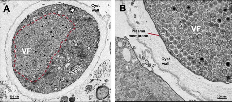
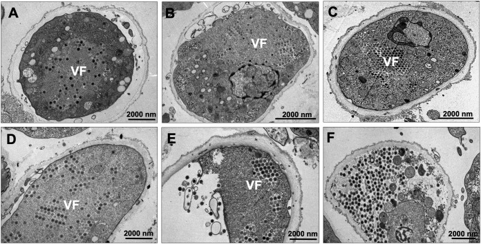
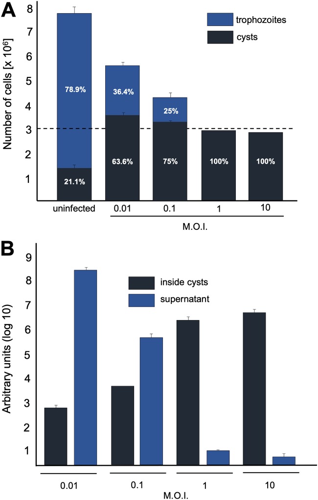
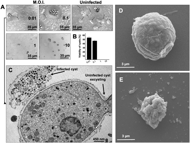
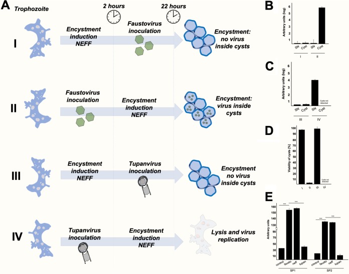
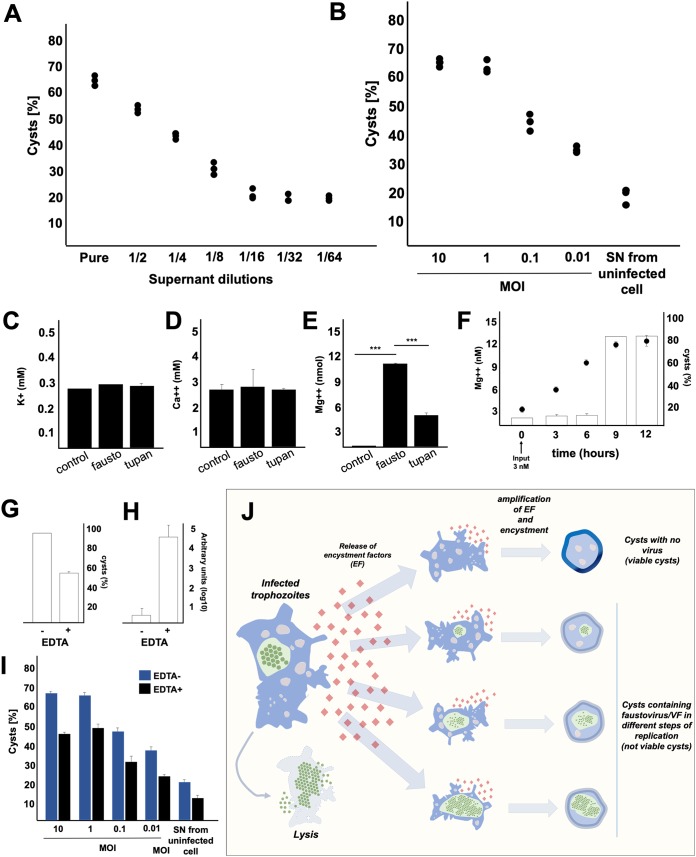
Viruses depend on cells to replicate and can cause considerable damage to their hosts. However, hosts have developed a plethora of antiviral mechanisms to counterattack or prevent viral replication and to maintain homeostasis. Advantageous features are constantly being selected, affecting host-virus interactions and constituting a harsh race for supremacy in nature. Here, we describe a new antiviral mechanism unveiled by the interaction between a giant virus and its amoebal host. Faustovirus mariensis infects Vermamoeba vermiformis, a free-living amoeba, and induces cell lysis to disseminate into the environment. Once infected, the cells release a soluble factor that triggers the encystment of neighbor cells, preventing their infection. Remarkably, infected cells stimulated by the factor encyst and trap the viruses and viral factories inside cyst walls, which are no longer viable and cannot excyst. This unprecedented mechanism illustrates that a plethora of antiviral strategies remains to be discovered in nature.IMPORTANCE Understanding how viruses of microbes interact with its hosts is not only important from a basic scientific point of view but also for a better comprehension of the evolution of life. Studies involving large and giant viruses have revealed original and outstanding mechanisms concerning virus-host relationships. Here, we report a mechanism developed by Vermamoeba vermiformis, a free-living amoeba, to reduce Faustovirus mariensis dissemination. Once infected, V. vermiformis cells release a factor that induces the encystment of neighbor cells, preventing infection of further cells and/or trapping the viruses and viral factories inside the cyst walls. This phenomenon reinforces the need for more studies regarding large/giant viruses and their hosts.

Keywords: antiviral; cysts; faustovirus; vermoameba; virus control.


Figures

FIG 1
FIG 1
Faustovirus mariensis isolation sites, particle images, and cytopathic effects. (A) Pampulha Lagoon map with collection sites highlighted (dots). The yellow dot represents where F. mariensis was collected, in front of the Pampulha Art Museum (top right of photo). Map courtesy of Google Maps. (B to D) F. mariensis viral particles visualized by scanning (B and C) and transmission (D) electron microscopy. (E to G) Plaque-forming unit (PFU) induced by F. mariensis infection in a Vermamoeba vermiformis monolayer. (F) Closeup of a PFU shown in panel E, observed 24 h postinfection. (G) Forty-eight hours postinfection, the PFUs expand and coalesce.
FIG 2
FIG 2
Gene categories and phylogeny. (A) F. mariensis gene set classified according to predicted gene categories. (B) DNA polymerase subunit B tree, constructed using the maximum likelihood evolution method and 1,000 replicates. Faustovirus mariensis (pentagon) clusters with other Faustovirus strains. Tree scale represents evolutionary distance.
FIG 3
FIG 3
Electron-lucent viral factory and cytoplasmic modifications induced by Faustovirus mariensis. (A to C) F. mariensis presents an electron-lucent viral factory (contoured in red and shown in detail in panel B), which was not easily distinguished from the rest of the cytoplasm and was observed at the perinuclear region. It is possible to visualize the abundant presence of mitochondria surrounding the viral factory (purple highlights in panels A and C). VF, viral factory; N, nucleus. Image B was obtained by TEM and graphically highlighted by using IOS image visualization software (Apple Technology Company).
FIG 4
FIG 4
Faustovirus mariensis morphogenesis and particle organization in honeycomb-like structures. (A) F. mariensis morphogenesis begins with crescents, open structures of approximately 50 nm, which grow as an electron-dense material of the viral factory fills them. Particles of almost 200 nm without genomic content are observed in late phases of morphogenesis, when the genome is incorporated and centralized within several newly formed viral particles. (B to E) F. mariensis progeny are organized in a honeycomb fashion inside viral factories. Small honeycombs expand as new mature viruses are formed and coalesce to others in the cytoplasm (B).
FIG 5
FIG 5
Faustovirus mariensis particles and viral factories inside Vermamoeba vermiformis cysts. (A) Mature V. vermiformis cyst enclosing a large viral factory (highlighted with a red dashed line). (B) Plasma membrane is clearly visible below the thick cyst wall. For this experiment, V. vermiformis cells were infected with F. mariensis at an MOI of 10 and prepared for transmission electron microscopy (TEM; 24 hpi).
FIG 6
FIG 6
Vermamoeba vermiformis cysts enclosing Faustovirus mariensis in different stages of viral replication cycle (A to F). The observation of more than 100 cysts revealed the presence of F. mariensis during distinct phases of the replication cycle, including early viral factory formation (A and B), late morphogenesis/honeycomb formation/coalescence (C to E), and cytoplasm filled with mature viral particles (F).
FIG 7
FIG 7
Cyst formation and viral replication at different multiplicities of infection (MOI). (A) Cyst and trophozoite quantification 48 h after the inoculation of F. mariensis at MOIs of 0.01, 0.1, 1, and 10. The dashed line represents the input of amoebae in the beginning of the experiment (3 × 106). Uninfected cells had a natural encystment rate of 21.1%. (B) F. mariensis genome quantification in the supernatants and cysts of V. vermiformis inoculated at different MOIs 1 week postinoculation. The supernatants and cysts were separated by centrifugation, and the viral genome load was quantified by qPCR (DNA polymerase subunit B). Error bars indicate standard deviations (SDs). These experiments were performed three times in triplicate.
FIG 8
FIG 8
Excystment assays. Cysts were produced by the inoculation of Vermamoeba vermiformis trophozoites with Faustovirus mariensis at MOIs of 0.01, 0.1, 1, and 10, and then their excystment potential was evaluated. (A) Agar plate excystment assay demonstrating that few cysts obtained from infections at MOIs of 0.01 and 0.1 were able to become trophozoites (arrows). Excystment was not observed for cysts obtained from infections at MOIs of 1 and 10. (B) Viability of cysts produced from infections at different MOIs (trypan blue, 0.4%). Error bars indicate SDs. (C) Transmission electron microscopy representative image demonstrating that only uninfected cysts are able to excyst. This image corresponds to cysts obtained from infections at an MOI of 0.01, after excystment stimulus. (D and E) Cysts produced after infection can present either a regular shape (D) or reduced diameters and irregular shapes (E). These experiments were performed three times in triplicate.
FIG 9
FIG 9
Faustovirus mariensis is not able to circumvent Vermamoeba vermiformis encystment. (A) Scheme highlighting the experimental strategy and results of an experiment testing the ability of F. mariensis and Tupanvirus to circumvent V. vermiformis encystment. In I and III, the encystment solution Neff was added prior to virus inoculation. In II and IV, the viruses were inoculated before Neff addition. (B) F. mariensis and (C) Tupanvirus genome quantification, in supernatant and cysts, for each experimental scenario analyzed (I to IV), by qPCR. The relative quantification was performed with the ΔCT method, and results were presented as arbitrary units (log10). (D) Quantification of the viability of cysts (0.4% trypan blue) produced from infections under different scenarios (I to IV). (E) Relative quantification of the expression of two serine proteinase mRNA isotypes present in V. vermiformis upon infection (MOI of 10) with F. mariensis or Tupanvirus or upon Neff treatment. The quantification was performed 5 h postinfection or after Neff inoculation. Error bars indicate SDs. The statistical significance was calculated using a two-tailed 2-way analysis of variance (ANOVA) test and Tukey’s range test, using GraphPad Prism. ***, P < 0.001. These experiments were performed three times in triplicate.
FIG 10
FIG 10
Investigation of encystment factors secreted by Vermamoeba vermiformis. (A) Induction of encystment (%) in V. vermiformis cells caused by the virus-free supernatant from a previous F. mariensis infection of a V. vermiformis culture (MOI of 10). Supernatant was diluted 2-fold from undiluted to 1/64. (B) Induction of encystment (%) of V. vermiformis cells caused by undiluted (pure) supernatant from a previous F. mariensis infection in V. vermiformis culture at different MOIs. (C to E) Quantification of the concentration of (C) K+, (D) Ca2+, and (E) Mg2+ in the supernatants of cells infected (MOI of 10, 10 hpi) with F. mariensis or Tupanvirus and in those of uninfected cells (control). The statistical significance was calculated using a two-tailed 2-way ANOVA test and a Tukey’s range test, using GraphPad Prism. ***, P < 0.001. (F) Evaluation of the potential of Mg2+ as an inductor of encystment in V. vermiformis. A total of 3 nmol of Mg2+ was added to a culture of 4 × 104 amoeba trophozoites, and the concentration of Mg2+ was measured, as well as the rate of cyst formation over time. (G and H) Evaluation of EDTA effect on (G) cyst formation and (H) viral genome replication during F. mariensis infection at an MOI of 10. (I) Evaluation of the inhibitory activity of 10 mM EDTA on the encystment process induced by supernatants of V. vermiformis infected by F. mariensis at different MOIs, 24 h postinoculation. For all graphs, error bars indicate SDs. These experiments were performed three times in triplicate. (J) Scheme summarizing the phenomenon described in this work. Once infected by F. mariensis, a V. vermiformis trophozoite can be lysed, as described for other amoebal giant viruses. However, during the first events of infection in a V. vermiformis population, encystment factors are released into the supernatant, which can induce the encystment of uninfected cells or even cause the encystment of infected trophozoites. As a result, we found cysts with no excystment ability during different stages of the replication cycle. The overall viral load in the supernatant is controlled by V. vermiformis trapping a substantial amount of viral progeny inside cysts.

Similar articles

Cited by

References

    1. Doron S, Melamed S, Ofir G, Leavitt A, Lopatina A, Keren M, Amitai G, Sorek R. 2018. Systematic discovery of antiphage defense systems in the microbial pangenome. Science 359:eaar4120. doi:10.1126/science.aar4120. - DOI - PMC - PubMed
    1. Ratcliff F, Harrison BD, Baulcombe DC. 1997. A similarity between viral defense and gene silencing in plants. Science 276:1558–1560. doi:10.1126/science.276.5318.1558. - DOI - PubMed
    1. Brubaker SW, Bonham KS, Zanoni I, Kagan JC. 2015. Innate immune pattern recognition: a cell biological perspective. Annu Rev Immunol 33:257–290. doi:10.1146/annurev-immunol-032414-112240. - DOI - PMC - PubMed
    1. Secombes CJ, Zou J. 2017. Evolution of interferons and interferon receptors. Front Immunol 8:209. - PMC - PubMed
    1. Borden EC, Sen GC, Uze G, Silverman RH, Ransohoff RM, Foster GR, Stark GR. 2007. Interferons at age 50: past, current and future impact on biomedicine. Nat Rev Drug Discov 6:975–990. doi:10.1038/nrd2422. - DOI - PMC - PubMed

Publication types

MeSH terms

LinkOut - more resources