. 2019 May 7;116(19):9333-9339.
doi: 10.1073/pnas.1814999116. Epub 2019 Apr 24.

The crystal structure of dGTPase reveals the molecular basis of dGTP selectivity

Affiliations

The crystal structure of dGTPase reveals the molecular basis of dGTP selectivity

Christopher O Barnes et al. Proc Natl Acad Sci U S A. .

Abstract

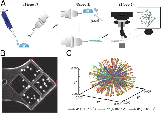
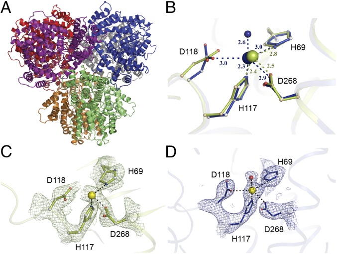
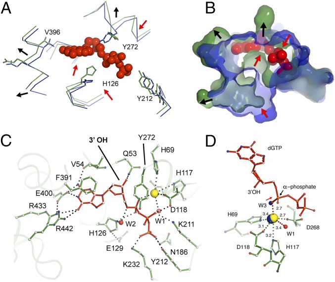
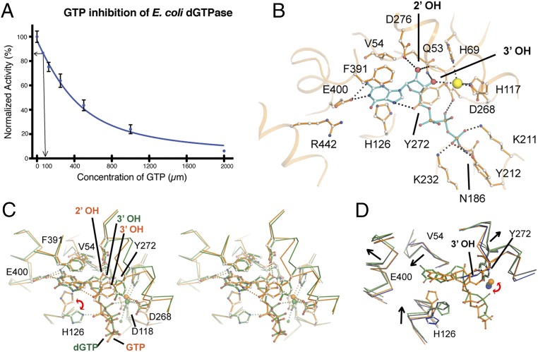
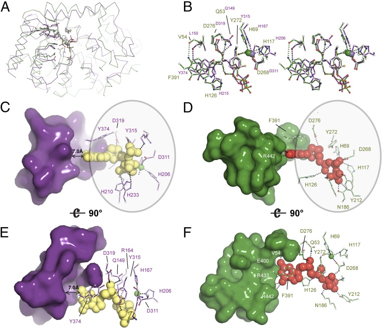
Deoxynucleotide triphosphohydrolases (dNTPases) play a critical role in cellular survival and DNA replication through the proper maintenance of cellular dNTP pools. While the vast majority of these enzymes display broad activity toward canonical dNTPs, such as the dNTPase SAMHD1 that blocks reverse transcription of retroviruses in macrophages by maintaining dNTP pools at low levels, Escherichia coli (Ec)-dGTPase is the only known enzyme that specifically hydrolyzes dGTP. However, the mechanism behind dGTP selectivity is unclear. Here we present the free-, ligand (dGTP)- and inhibitor (GTP)-bound structures of hexameric Ec-dGTPase, including an X-ray free-electron laser structure of the free Ec-dGTPase enzyme to 3.2 Å. To obtain this structure, we developed a method that applied UV-fluorescence microscopy, video analysis, and highly automated goniometer-based instrumentation to map and rapidly position individual crystals randomly located on fixed target holders, resulting in the highest indexing rates observed for a serial femtosecond crystallography experiment. Our structures show a highly dynamic active site where conformational changes are coupled to substrate (dGTP), but not inhibitor binding, since GTP locks dGTPase in its apo- form. Moreover, despite no sequence homology, Ec-dGTPase and SAMHD1 share similar active-site and HD motif architectures; however, Ec-dGTPase residues at the end of the substrate-binding pocket mimic Watson-Crick interactions providing guanine base specificity, while a 7-Å cleft separates SAMHD1 residues from dNTP bases, abolishing nucleotide-type discrimination. Furthermore, the structures shed light on the mechanism by which long distance binding (25 Å) of single-stranded DNA in an allosteric site primes the active site by conformationally "opening" a tyrosine gate allowing enhanced substrate binding.

Keywords: X-ray free-electron laser; dNTP triphosphohydrolase; metalloenzymes; serial femtosecond crystallography.


Conflict of interest statement

The authors declare no conflict of interest.

Figures

Fig. 1.
Fig. 1.
Loading and mapping of crystals on MCHs. (A) Schematic representation of MCH loading as detailed in Methods. (B) UV-microscopy image of Pol-Spt4/5 mounted crystals. Fiducial marks are indicated by a red asterisk. (C) Reciprocal space representation of the basis vectors of 221 indexed dGTPase images demonstrating the lack of preferential alignment when mounting in MCHs.
Fig. 2.
Fig. 2.
The hexameric Ec-dGTPase XFEL crystal structure. (A) Cartoon representation of the hexameric Ec-dGTPase XFEL crystal structure solved using 221 still images from randomly oriented crystals. (B) Differences in Mn2+ coordination for XFEL (blue) and low-dose synchrotron structure (olive green). Potential H-bond interactions with distances shorter than 3.5 Å are indicated as dashes between residues. (C) Electron density of the Sigma-A weighted 2FobsFcalc map contoured at 1.5σ for residues comprising the HD motif (synchrotron data); Mn2+ ion is illustrated as a yellow sphere. (D) Electron density of the Sigma-A weighted 2FobsFcalc map contoured at 1.5σ for residues comprising the HD motif (XFEL data). A water molecule (indicated in red) forms part of the Mn2+ coordination (SI Appendix, Fig. S2C).
Fig. 3.
Fig. 3.
Interactions of dGTP substrate with Ec-dGTPase. Wire (A) and surface (B) representations of the overlay between the apo- (blue) and dGTP-bound (green) structures showing the conformational changes observed in the active-site pocket upon dGTP binding (red spheres). Red arrows indicate contraction, and black arrows indicate expansion of the pocket. (C) Ball-and-stick representation of key residues (green) involved in dGTP (red) binding. Hydrogen bonds are illustrated as dashes and water molecules as red spheres. (D) Ball-and-stick representation of Mn2+ (yellow sphere) coordination by dGTP residues (green). Distances are indicated next to dashes. The position of the apo-XFEL Mn2+ ion and coordinating water after overlay with the dGTP bound-structure are indicated as blue spheres.
Fig. 4.
Fig. 4.
Inhibition of Ec-dGTPase activity by GTP. (A) Activity of Ec-dGTPase in the presence of increasing concentrations of GTP (µM) and 100 µM dGTP substrate. Enzymatic activity assays were repeated three times, and the SD was plotted (n = 3). The arrows indicate dGTPase activity in the presence of 100 µM GTP. (B) Ball-and-stick representation and potential hydrogen bond interactions (black dashes) between GTP (cyan) and active-site residues of dGTPase (orange). Asp276 and Gln53 form H-bonds with the 2′-OH and the 3′-OH, respectively. Coordination of the metal by an oxygen from the α-phosphate in the dGTP-bound form is swapped by the 3′-OH of the ribose, resulting in 1.5 Å displacement with respect to its position in the dGTP-bound form; as result of this positional change, HD motif residue Asp118 no longer forms part of its coordination sphere. (C) Stereo and ball-and-stick representations of the overlay between the dGTP- (green) and the GTP-bound (orange) structures illustrating that the two binding pockets differ significantly. (D) Overlay of the substrate-bound structures (dGTP and dGTP-1-thiol, green and black, respectively) with the XFEL-apo and GTP-bound (inhibited) structures (blue and orange) illustrating that GTP binding “locks” the active site hindering the conformational changes observed during substrate binding. The rmsd differences between active-site residues for XFEL, dGTP, dGTP-1-thiol, and GTP are summarized in SI Appendix, Table S3.
Fig. 5.
Fig. 5.
Structural and enzymatic insight into the mechanism of Ec-dGTPase activity. (A) Ribbon representation of the overlay between SAMHD1 (PDB ID code 4BZC) (purple) and Ec-dGTPase (green) illustrating fold conservation of the enzymatic cores. (B) Stereo and ball-and-stick representation of the overlay between SAMHD1 and Ec-dGTPase active sites illustrating residue type and geometry conservation. (C and E) Surface and ball-and-stick representation of SAMHD1 active-site residues illustrating that most contacts with the dNTP involve interaction with the ribose, the phosphates, and the purine or pyrimidine ring (circle). No interactions with SAMHD1 residues that could confer specificity are possible since a 7-Å gap separates them from the dNTP. Thus, dNTPases bind shared motifs D and F. A similar set of interactions takes place in dGTPase; however, dGTP selectivity occurs through formation of four hydrogen bonds.

Similar articles

Cited by

References

    1. Meuth M. The genetic consequences of nucleotide precursor pool imbalance in mammalian cells. Mutat Res. 1984;126:107–112. - PubMed
    1. Seto D, Bhatnagar SK, Bessman MJ. The purification and properties of deoxyguanosine triphosphate triphosphohydrolase from Escherichia coli. J Biol Chem. 1988;263:1494–1499. - PubMed
    1. Nordlund P, Reichard P. Ribonucleotide reductases. Annu Rev Biochem. 2006;75:681–706. - PubMed
    1. Kunz BA, et al. International commission for protection against environmental mutagens and carcinogens. Deoxyribonucleoside triphosphate levels: A critical factor in the maintenance of genetic stability. Mutat Res. 1994;318:1–64. - PubMed
    1. Wheeler LJ, Rajagopal I, Mathews CK. Stimulation of mutagenesis by proportional deoxyribonucleoside triphosphate accumulation in Escherichia coli. DNA Repair (Amst) 2005;4:1450–1456. - PubMed

Publication types

MeSH terms

Substances