. 2019 May 7;116(19):9443-9452.
doi: 10.1073/pnas.1814097116. Epub 2019 Apr 24.

Human skin long noncoding RNA WAKMAR1 regulates wound healing by enhancing keratinocyte migration

Affiliations

Human skin long noncoding RNA WAKMAR1 regulates wound healing by enhancing keratinocyte migration

Dongqing Li et al. Proc Natl Acad Sci U S A. .

Abstract

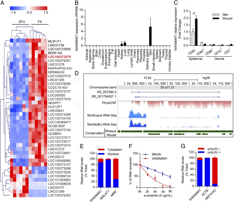
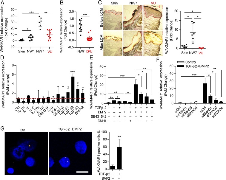
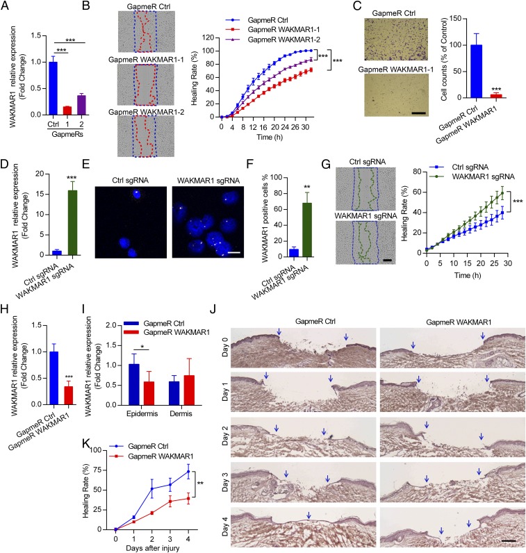
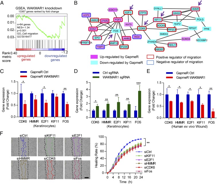
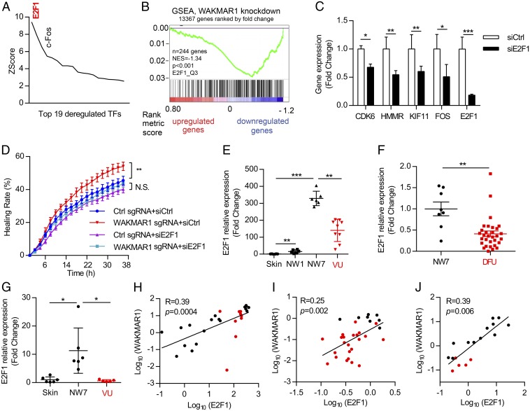
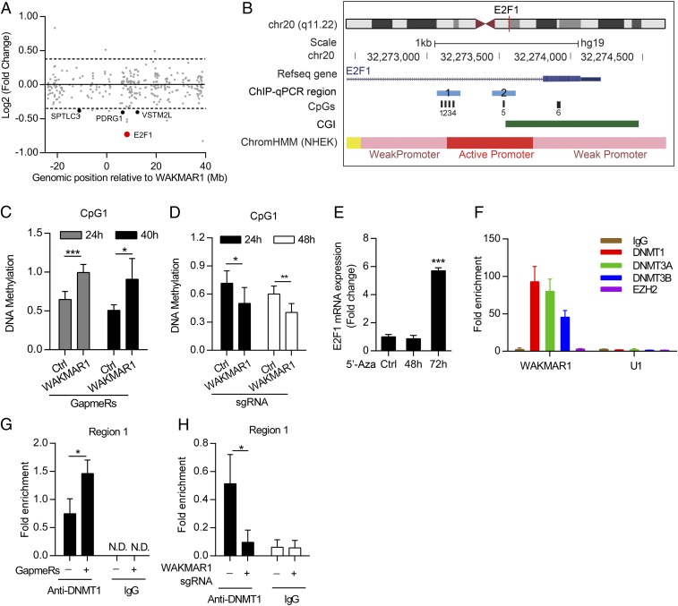
An increasing number of studies reveal the importance of long noncoding RNAs (lncRNAs) in gene expression control underlying many physiological and pathological processes. However, their role in skin wound healing remains poorly understood. Our study focused on a skin-specific lncRNA, LOC105372576, whose expression was increased during physiological wound healing. In human nonhealing wounds, however, its level was significantly lower compared with normal wounds under reepithelialization. We characterized LOC105372576 as a nuclear-localized, RNAPII-transcribed, and polyadenylated lncRNA. In keratinocytes, its expression was induced by TGF-β signaling. Knockdown of LOC105372576 and activation of its endogenous transcription, respectively, reduced and increased the motility of keratinocytes and reepithelialization of human ex vivo skin wounds. Therefore, LOC105372576 was termed "wound and keratinocyte migration-associated lncRNA 1" (WAKMAR1). Further study revealed that WAKMAR1 regulated a network of protein-coding genes important for cell migration, most of which were under the control of transcription factor E2F1. Mechanistically, WAKMAR1 enhanced E2F1 expression by interfering with E2F1 promoter methylation through the sequestration of DNA methyltransferases. Collectively, we have identified a lncRNA important for keratinocyte migration, whose deficiency may be involved in the pathogenesis of chronic wounds.

Keywords: keratinocyte migration; long noncoding RNA; wound healing.


Conflict of interest statement

The authors declare no conflict of interest.

Figures

Fig. 1.
Fig. 1.
WAKMAR1 is a nuclear-localized, RNAPII-transcribed, and polyadenylated lncRNA. (A) Heat map illustrates the differentially expressed lncRNAs (absolute fold change ≥ 3, false discovery rate < 0.01) in DFU compared with foot skin (FS). LOC105372576/WAKMAR1 is highlighted in red. Z-score transformation was applied for visualization. Data are from a published microarray dataset (GEO accession no. GSE80178; https://www.ncbi.nlm.nih.gov/geo/query/acc.cgi?acc=gse80178). (B) WAKMAR1 expression in human tissues. RPKM, reads per kilobase per million reads. (C) qPCR analysis of WAKMAR1 in epidermal CD45 and CD45+ cells and in dermal CD90+, CD14+, and CD3+ cells, which were isolated from the skin and day 7 wounds of healthy donors (n = 4). *P < 0.05, unpaired two-tailed Student’s t test. (D) Genomic snapshot of WAKMAR1 generated in RefSeq (purple), phylogenetic information-based codon substitution frequency (PhyloCSF; red), RNA-sequencing (blue) of human skin with (SkinExpos) or without sun exposure (SkinNotEx), and conservation (green) tracks. (E) qPCR analysis of WAKMAR1, MALAT1, and HPRT in the nucleus or cytoplasm of keratinocytes (n = 3). (F) qPCR analysis of WAKMAR1 and RPLP0 mRNA in keratinocytes treated with α-amanitin (n = 3). (G) qPCR analysis of WAKMAR1, ACTB, and HIS1H1D in Poly(A)+ and Poly(A) RNA fractions from keratinocytes (n = 3). Data are presented as mean ± SEM (B and C) or mean ± SD (EG).
Fig. 2.
Fig. 2.
WAKMAR1 expression is decreased in wound-edge keratinocytes of human chronic wounds and regulated by TGF-β. (A) qPCR analysis of WAKMAR1 in the skin, in day 1 (NW1) and day 7 (NW7) normal wounds from six healthy donors, and in wound edges of nine patients with VU. (B) qPCR analysis of WAKMAR1 in NW7 biopsies from eight healthy donors and wound edges of 29 patients with DFU. (C) qPCR analysis of WAKMAR1 in wound-edge epidermis isolated from the healthy skin and NW7 (n = 6) and VU (n = 5) using LCM. (Scale bar, 200 μm.) Red arrows indicate wound edges. (D) qPCR analysis of WAKMAR1 in keratinocytes treated with wound-related cytokines/growth factors for 24 h (n = 3). Ctrl, control. (E) TGF-β receptor inhibitor SB431542 and/or BMP receptor inhibitor DMH1 was applied 15 min before adding TGF-β2 and/or BMP2 to keratinocytes, and WAKMAR1 was analyzed by qPCR 24 h later (n = 3). (F) qPCR analysis of WAKMAR1 in keratinocytes transfected with SMAD1-, SMAD3-, and SMAD4-specific siRNAs for 24 h and then treated with TGF-β2 and BMP2 for 24 h (n = 3). (G) ISH of WAKMAR1 in keratinocytes treated with TGF-β2 and BMP2 for 24 h. Cell nuclei were costained with DAPI. (Scale bar, 50 μm.) WAKMAR1+ cells were counted. *P < 0.05; **P < 0.01; ***P < 0.001 by Mann–Whitney U test (AC) and unpaired two-tailed Student’s t test (DG). Data are presented as mean ± SEM (AC) or mean ± SD (DG).
Fig. 3.
Fig. 3.
WAKMAR1 regulates keratinocyte motility and wound reepithelialization. (A) qPCR analysis of WAKMAR1 in keratinocytes transfected with WAKMAR1-specific GapmeR1, GapmeR2, or control oligos (Ctrl) (n = 3). (B) Scratch wound assay of keratinocytes after WAKMAR1 knockdown (n = 10). (C) Representative photographs of transwell migration assay for keratinocytes with WAKMAR1 knockdown (n = 3). (Scale bar, 1 mm.) The number of cells passing through the membrane was counted. qPCR (D) and ISH (E and F) of WAKMAR1 in keratinocytes transfected with CRISPR/Cas9-SAM plasmids for 48 h are shown. Cell nuclei were costained with DAPI. (Scale bar, 50 μm.) (G) Scratch wound assay of keratinocytes with WAKMAR1 expression activation (n = 10). (Scale bar, 300 μm.) qPCR analyses of WAKMAR1 in full-depth biopsies (H) and in LCM-isolated epidermis and dermis (I) of human ex vivo wounds after topical application of WAKMAR1 GapmeRs for 4 d (n = 6 donors) are shown. (J) Representative photographs of hematoxylin and eosin staining of ex vivo wounds. Blue arrows demarcate the initial wound edges (day 0) and newly formed epidermis (days 1–4). (Scale bar, 200 μm.) (K) Reepithelialization was quantified as healing rate = 100% − percentage of the initial wound size. *P < 0.05; **P < 0.01; ***P < 0.001 by unpaired two-tailed Student’s t test (A, C, D, F, H, and I) or two way-ANOVA (B, G, and K). Data are presented as mean ± SD and are representative of at least two independent experiments.
Fig. 4.
Fig. 4.
WAKMAR1 regulates a gene network mediating its promigratory function in keratinocytes. Microarray analysis was performed in human keratinocytes with WAKMAR1 knockdown (n = 3). (A) GSEA evaluated enrichment for the cell migration-related genes in the microarray data. NES, normalized enrichment score. (B) Functional protein association network was identified by STRING APP in Cytoscape software among the genes regulated by WAKMAR1 (absolute fold change ≥ 1.3, P < 0.05). Genes up- or down-regulated by WAKMAR1 GapmeR are colored in pink or cyan, respectively. Genes previously reported to promote or inhibit cell migration are highlighted with red or blue frames, respectively. The expression of CDK6, HMMR, E2F1, KIF11, and FOS was analyzed by qPCR in keratinocytes transfected with WAKMAR1 GapmeRs (C), or with CRISPR/Cas9-SAM plasmids (D), and in human ex vivo wounds treated with WAKMAR1 GapmeRs (E). (F) Scratch wound assay of keratinocytes transfected with siRNAs specific to KIF11, E2F1, HMMR, CDK6, or FOS (n = 8). (Scale bar, 300 μm.) *P < 0.05; **P < 0.01; ***P < 0.001 by unpaired two-tailed Student’s t test (CE) or two way-ANOVA (F). Data are presented as mean ± SD and are representative of at least two independent experiments.
Fig. 5.
Fig. 5.
E2F1 acts as an upstream regulator in the WAKMAR1-regulated gene network. (A) MetaCore analysis identified transcription factors (TFs) with overrepresented binding sites among WAKMAR1-regulated genes. Only TFs with changed expression after WAKMAR1 knockdown are shown. (B) GSEA evaluated the enrichment of E2F1-target genes in the WAKMAR1-regulated genes. NES, normalized enrichment score. (C) qPCR analysis of CDK6, HMMR, KIF11, FOS, and E2F1 in keratinocytes transfected with E2F1 siRNA for 24 h (n = 3). (D) Scratch wound assay of keratinocytes after WAKMAR1 knockdown and/or E2F1 silencing (n = 8). qPCR analyses of E2F1 in the skin in day 1 (NW1) and day 7 (NW7) normal wounds from six healthy donors and in the wound edges of nine patients with VU (E and H) in NW7 (n = 8) vs. DFU (n = 29) (F and I) and in wound-edge epidermis isolated from healthy skin and NW7 (n = 6) and VU (n = 5) with LCM (G and J) are shown. (HJ) Expression correlation of WAKMAR1 with E2F1 in human skin, normal wounds (black dots), and chronic wounds (red dots). *P < 0.05; **P < 0.01; ***P < 0.001 by unpaired two-tailed Student’s t test (C), two way-ANOVA (D), Mann–Whitney U test (EG), or Pearson’s correlation test (HJ). N.S., not significant. Data are presented as mean ± SD (C) or mean ± SEM (DG).
Fig. 6.
Fig. 6.
WAKMAR1 activates E2F1 expression by suppressing E2F1 promoter methylation. (A) Chr20 gene expression in keratinocytes with WAKMAR1 knockdown. The gene start distance from the WAKMAR1 transcriptional start site is shown on the x axis. The log-twofold change of expression levels between control and WAKMAR1 knockdown is shown on the y axis. Genes with absolute fold change ≥ 1.3 and P < 0.05 are highlighted. (B) Genomic snapshot of E2F1 promoter: CpG sites (black bars) analyzed by MSRE-qPCR (CpG1, CpG5, and CpG6) and bisulfite pyrosequencing (CpG1, CpG2, CpG3, and CpG4) and regions analyzed by ChIP-qPCR (blue bars) are highlighted. CGI, CpG island; ChromHMM, chromatin state segmentation by hidden Markov model from ENCODE/Broad; NHEK, normal human epidermal keratinocytes. MSRE-qPCR analyses of DNA methylation at CpG1 in keratinocytes transfected with WAKMAR1 GapmeR for 24 h and 40 h (n = 6) (C) or CRISPR/Cas9-SAM plasmids for 24 h and 48 h (n = 6) (D) are shown. Ctrl, control. (E) qPCR of E2F1 in keratinocytes treated with 5′-Aza-2′-deoxycytidine (5′-Aza) (n = 3). (F) qPCR analysis of WAKMAR1 and U1 small nuclear RNA immunoprecipitated from keratinocytes with DNMT1, DNMT3A, DNMT3B, and EZH2 antibodies or IgG (n = 3). ChIP-qPCR of E2F1 promoter region 1 was performed in keratinocytes transfected with WAKMAR1 GapmeRs (G) or CRISPR/Cas9-SAM plasmids (H) and immunoprecipitated using DNMT1 antibody or IgG (n = 3). *P < 0.05; ***P < 0.001 by unpaired two-tailed Student’s t test (CE, G, and H). N.D., not detected. Data are presented as mean ± SD.

References

    1. Reinke JM, Sorg H. Wound repair and regeneration. Eur Surg Res. 2012;49:35–43. - PubMed
    1. Eming SA, Martin P, Tomic-Canic M. Wound repair and regeneration: Mechanisms, signaling, and translation. Sci Transl Med. 2014;6:265sr6. - PMC - PubMed
    1. Kopp F, Mendell JT. Functional classification and experimental dissection of long noncoding RNAs. Cell. 2018;172:393–407. - PMC - PubMed
    1. Volders PJ, et al. An update on LNCipedia: A database for annotated human lncRNA sequences. Nucleic Acids Res. 2015;43:4363–4364. - PMC - PubMed
    1. Liu SJ, et al. CRISPRi-based genome-scale identification of functional long noncoding RNA loci in human cells. Science. 2017;355:aah7111. - PMC - PubMed

Publication types