

Helical nanofiber yarn enabling highly stretchable engineered microtissue
- PMID: 31019088
- PMCID: PMC6511022
- DOI: 10.1073/pnas.1821617116
Helical nanofiber yarn enabling highly stretchable engineered microtissue
Abstract
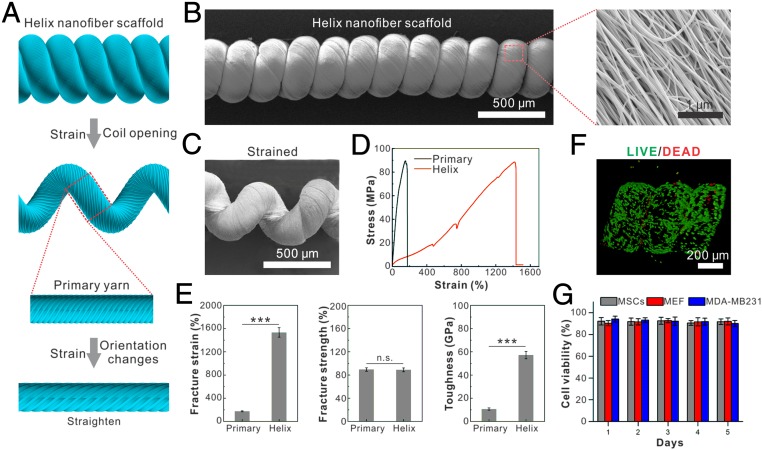
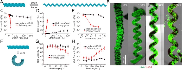
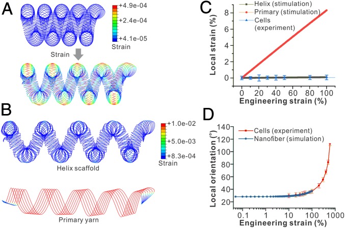
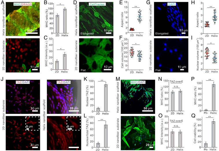
Development of microtissues that possess mechanical properties mimicking those of native stretchable tissues, such as muscle and tendon, is in high demand for tissue engineering and regenerative medicine. However, regardless of the significant advances in synthetic biomaterials, it remains challenging to fabricate living microtissue with high stretchability because application of large strains to microtissues can damage the cells by rupturing their structures. Inspired by the hierarchical helical structure of native fibrous tissues and its behavior of nonaffine deformation, we develop a highly stretchable and tough microtissue fiber made up of a hierarchical helix yarn scaffold, scaling from nanometers to millimeters, that can overcome this limitation. This microtissue can be stretched up to 15 times its initial length and has a toughness of 57 GJ m-3 More importantly, cells grown on this scaffold maintain high viability, even under severe cyclic strains (up to 600%) that can be attributed to the nonaffine deformation under large strains, mimicking native biopolymer scaffolds. Furthermore, as proof of principle, we demonstrate that the nanotopography of the helical nanofiber yarn is able to induce cytoskeletal alignment and nuclear elongation, which promote myogenic differentiation of mesenchymal stem cells by triggering nuclear translocation of transcriptional coactivator with PDZ-binding motif (TAZ). The highly stretchable microtissues we develop here will facilitate a variety of tissue engineering applications and the development of engineered living systems.
Keywords: bioinspired scaffold; muscle regeneration; myogenesis; nanofiber yarn; stretchable tissue.
Conflict of interest statement
The authors declare no conflict of interest.
Figures
Similar articles
-
Combining a micro/nano-hierarchical scaffold with cell-printing of myoblasts induces cell alignment and differentiation favorable to skeletal muscle tissue regeneration.Biofabrication. 2016 Sep 16;8(3):035021. doi: 10.1088/1758-5090/8/3/035021. Biofabrication. 2016. PMID: 27634918
-
Mesenchymal stem cells and myoblast differentiation under HGF and IGF-1 stimulation for 3D skeletal muscle tissue engineering.BMC Cell Biol. 2017 Feb 28;18(1):15. doi: 10.1186/s12860-017-0131-2. BMC Cell Biol. 2017. PMID: 28245809 Free PMC article.
-
Nanofiber Yarn/Hydrogel Core-Shell Scaffolds Mimicking Native Skeletal Muscle Tissue for Guiding 3D Myoblast Alignment, Elongation, and Differentiation.ACS Nano. 2015 Sep 22;9(9):9167-79. doi: 10.1021/acsnano.5b03644. Epub 2015 Aug 19. ACS Nano. 2015. PMID: 26280983
-
Electrospun three-dimensional aligned nanofibrous scaffolds for tissue engineering.Mater Sci Eng C Mater Biol Appl. 2018 Nov 1;92:995-1005. doi: 10.1016/j.msec.2018.06.065. Epub 2018 Jun 30. Mater Sci Eng C Mater Biol Appl. 2018. PMID: 30184829 Review.
-
New state of nanofibers in regenerative medicine.Artif Cells Nanomed Biotechnol. 2017 Mar;45(2):204-210. doi: 10.3109/21691401.2016.1170696. Epub 2016 Apr 13. Artif Cells Nanomed Biotechnol. 2017. PMID: 27075144 Review.
Cited by
-
Ion-Specific Assembly of Strong, Tough, and Stiff Biofibers.Angew Chem Int Ed Engl. 2019 Dec 16;58(51):18562-18569. doi: 10.1002/anie.201910603. Epub 2019 Nov 4. Angew Chem Int Ed Engl. 2019. PMID: 31600016 Free PMC article. Review.
-
State-of-the-art review of advanced electrospun nanofiber yarn-based textiles for biomedical applications.Appl Mater Today. 2022 Jun;27:101473. doi: 10.1016/j.apmt.2022.101473. Epub 2022 Apr 10. Appl Mater Today. 2022. PMID: 35434263 Free PMC article. Review.
-
How Advancing is Peripheral Nerve Regeneration Using Nanofiber Scaffolds? A Comprehensive Review of the Literature.Int J Nanomedicine. 2023 Nov 15;18:6763-6779. doi: 10.2147/IJN.S436871. eCollection 2023. Int J Nanomedicine. 2023. PMID: 38026517 Free PMC article. Review.
-
Morphological Hydrogel Microfibers with MXene Encapsulation for Electronic Skin.Research (Wash D C). 2021 Mar 3;2021:7065907. doi: 10.34133/2021/7065907. eCollection 2021. Research (Wash D C). 2021. PMID: 33763650 Free PMC article.
-
Dual-Responsive MXene-Functionalized Wool Yarn Artificial Muscles.Adv Sci (Weinh). 2024 Jul;11(25):e2402196. doi: 10.1002/advs.202402196. Epub 2024 Apr 22. Adv Sci (Weinh). 2024. PMID: 38650164 Free PMC article.
References
-
- Fung YC. Biomechanics: Mechanical Properties of Living Tissues. Springer Science & Business Media; New York: 2013.
-
- Place ES, Evans ND, Stevens MM. Complexity in biomaterials for tissue engineering. Nat Mater. 2009;8:457–470. - PubMed
-
- Gao G, Du G, Sun Y, Fu J. Self-healable, tough, and ultrastretchable nanocomposite hydrogels based on reversible polyacrylamide/montmorillonite adsorption. ACS Appl Mater Interfaces. 2015;7:5029–5037. - PubMed
Publication types
MeSH terms
Substances
LinkOut - more resources
Full Text Sources
Other Literature Sources
