. 2019 Apr 24;9(1):6516.
doi: 10.1038/s41598-019-42751-4.

Airway surface liquid acidification initiates host defense abnormalities in Cystic Fibrosis

Affiliations

Airway surface liquid acidification initiates host defense abnormalities in Cystic Fibrosis

Juliette Simonin et al. Sci Rep. .

Erratum in

Abstract

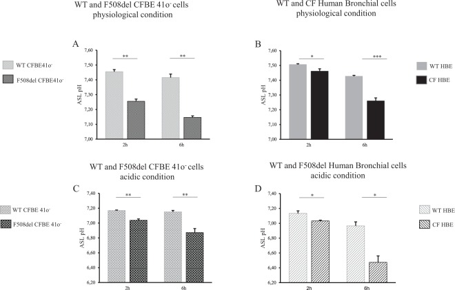
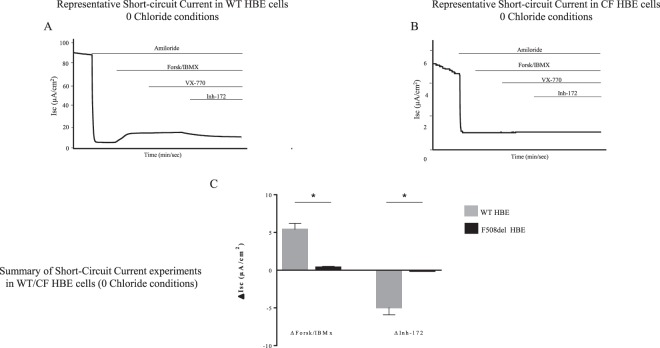
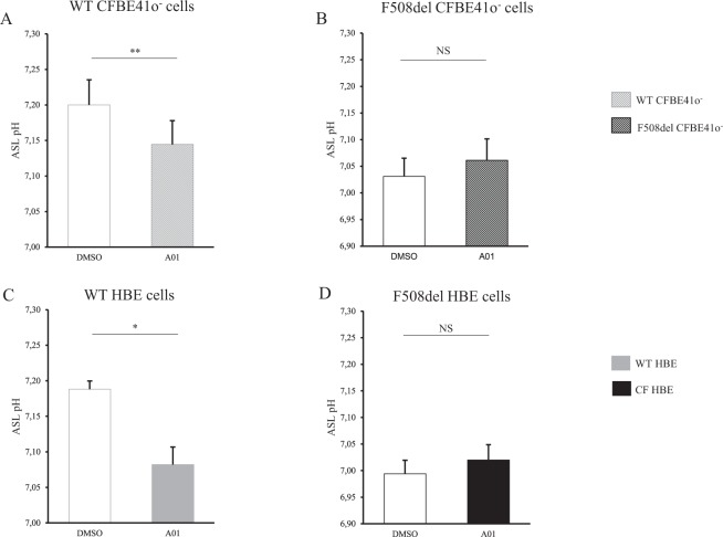
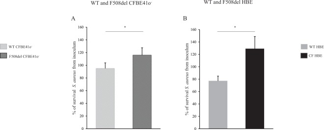
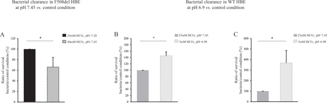
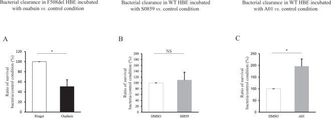
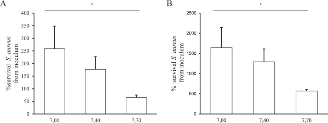
Cystic fibrosis (CF) is caused by defective Cystic Fibrosis Transmembrane Conductance Regulator (CFTR) protein. Morbidity is mainly due to early airway infection. We hypothesized that S. aureus clearance during the first hours of infection was impaired in CF human Airway Surface Liquid (ASL) because of a lowered pH. The ASL pH of human bronchial epithelial cell lines and primary respiratory cells from healthy controls (WT) and patients with CF was measured with a pH microelectrode. The antimicrobial capacity of airway cells was studied after S. aureus apical infection by counting surviving bacteria. ASL was significantly more acidic in CF than in WT respiratory cells. This was consistent with a defect in bicarbonate secretion involving CFTR and SLC26A4 (pendrin) and a persistent proton secretion by ATP12A. ASL demonstrated a defect in S. aureus clearance which was improved by pH normalization. Pendrin inhibition in WT airways recapitulated the CF airway defect and increased S. aureus proliferation. ATP12A inhibition by ouabain decreased bacterial proliferation. Antimicrobial peptides LL-37 and hBD1 demonstrated a pH-dependent activity. Normalizing ASL pH might improve innate airway defense in newborns with CF during onset of S. aureus infection. Pendrin activation and ATP12A inhibition could represent novel therapeutic strategies to normalize pH in CF airways.


Conflict of interest statement

The authors declare no competing interests.

Figures

Figure 1
Figure 1
Airway surface liquid pH measurements under physiological and normocapnic acidosis conditions in WT and F508del bronchial epithelia at 2 and 6 hours. Apical fluid pH was measured after apical addition for 2 hours or 6 hours of 50 µL of physiologic Ringer’s solution (25 mM HCO3, 5% CO2) (upper panel) or acidic Ringer’s (10 mM HCO3, 5% CO2) (lower panel). (A) WT vs. F508del CFBE41o cells. 2 h: pH = 7.45 ± 0.01 vs. pH = 7.26 ± 0.01, p = 0.008; 6 h: pH = 7.42 ± 0.02 vs. pH = 7.15 ± 0.01, p = 0.008. (B) WT vs. CF Human Bronchial Epithelial (HBE) primary cells. 2 h: pH = 7.51 ± 0.004 vs. pH = 7.46 ± 0.02, p = 0.036; 6 h: pH = 7.43 ± 0.006 vs. pH = 7.26 ± 0.02, p < 0.001. (C) WT vs. F508del CFBE41o cells. 2 h: pH = 7.17 ± 0.01 vs. pH = 7.04 ± 0.02, p = 0.008; 6 h: pH = 7.15 ± 0.02 vs. pH = 6.87 ± 0.05, p = 0.008. (D) WT vs. F508del HBE primary cells. 2 h: pH = 7.13 ± 0.03 vs. pH = 7.03 ± 0.01, p = 0.028; 6 h: pH = 6.97 ± 0.05 vs. pH = 6.47 ± 0.09, p = 0.028. For all conditions, n = 3 in triplicate except for figure B where CF bronchial primary cells were derived from 3 F508del homozygote donors (triplicate for each patient) and 1 N1303K/4005 + 1G > A patient (12 filters). Data are presented as mean ± SEM. Statistical significance from unpaired nonparametric Wilcoxon test. *p < 0.05; **p < 0.01; ***p < 0.001; NS: non significant.
Figure 2
Figure 2
Airway surface liquid pH measurements upon ATP12A, NBC1 and SLC26A4 activity inhibition in WT and F508del Human Bronchial Epithelial primary cells. pH changes (ΔpH) in Human Bronchial Epithelial (HBE) primary cells after incubation with vehicle or specific inhibitor are shown in WT HBE cells (grey bars) or F508del HBE (black bars). Apical fluid pH was measured after apical addition of 50 µL of physiologic Ringer’s solution (25 mM HCO3, 5% CO2) for 2 hours. Ouabain 1 mM (or Ringer as vehicle) was added at the apical side for 2 hours, as ATP12A inhibitor; S0859 100 µM (or DMSO as vehicle) was added at the basal side for 2 hours, as NBC1 inhibitor; A01, 25 µM (or DMSO as vehicle) was added at the basal side for 6 hours, as SLC26A4 inhibitor. Ouabain. WT HBE: ΔpH (Ouabain-Ringer) = 0.095 ± 0.009 (p = 0.002); F508del HBE: ΔpH = 0.09 ± 0.013 (p = 0.028); WT vs. F508del HBE ΔpH: NS. For all conditions, n = 4 in triplicate. S0859. WT HBE: ΔpH (S0859-DMSO) = −0.02 ± 0.014 (NS); F508del HBE: ΔpH = −0.02 ± 0.011 (NS); WT vs. F508del HBE ΔpH: NS. For all conditions, n = 3 in triplicate. A01. WT HBE ΔpH (A01-DMSO) = −0.10 ± 0.022 (p = 0.043); F508del HBE ΔpH = 0.068 ± 0.041 (NS); WT vs. F508del HBE ΔpH: p = 0.04. For all conditions, n = 3 in triplicate. Data are presented as mean ± SEM. Statistical significance from unpaired nonparametric Wilcoxon test. Level of significance is shown for ΔpH in each genotype as well as the comparison between genotypes. *p < 0.05; **p < 0.01; NS: non significant.
Figure 3
Figure 3
Short-circuit current measurements under 0 Chloride conditions in WT and CF Human Bronchial Epithelial primary cells. Experiments were done in a solution with 0 Cl and 25 mM HCO3, bubbled with 95% O2 and 5% CO2 at the apical and basal faces. Short-circuit current variation were induced by successive addition of amiloride (100 µM), Forskolin/IBMX (10 µM/100 µM), VX770 (10 µM), Inh-172 (5 µM). (A) Representative experiment from WT Human Bronchial Epithelial (HBE) primary cells. (B) Representative experiment from CF HBE primary cells (N1303K/4005 + 1G > A). (C) Summary of results obtained from 3 WT subjects (n = 3; 14 filters) (grey bars) and 1 CF patient (N1303K/4005 + 1G > A; 2 independent experiments; 7 filters) (black bars). ΔForsk/IBMX: 5.4 ± 0.8 µA/cm2 in WT HBE vs. 0.4 ± 0.1 /cm2 in CF HBE; p = 0.012. ΔInh-172: −4.9 ± 0.97 µA/cm2 in WT HBE vs. −0.1 ± 0.05 µA/cm2 in F508del HBE; p = 0.02. Data are presented as mean ± SEM. Statistical significance from unpaired nonparametric Wilcoxon test. *p < 0.05.
Figure 4
Figure 4
Airway surface liquid pH measurements upon SLC26A4 inhibition in WT and F508del bronchial epithelia. CFBE41o cells and Human Bronchial Epithelial (HBE) primary cells were incubated with DMSO vehicle (empty bars) or specific pendrin inhibitor A01 (filled bars) for 6 h. ASL pH was measured after apical addition of 50 µL Ringer’s solution (25 mM HCO3, 5% CO2). DMSO induces a slight ASL acidification in both WT and F508del cells reaching a decrease of 0.2 pH units after 6 h incubation. (A) WT CFBE41o cells. pH = 7.20 ± 0.04 (DMSO) vs. pH = 7.14 ± 0.03 (A01), p = 0.008. (B) F508del CFBE41o cells. pH = 7.03 ± 0.03 (DMSO) vs. pH = 7.06 ± 0.04 (A01), NS. (C) WT HBE primary cells. pH = 7.19 ± 0.01 (DMSO) vs. pH = 7.08 ± 0.02 (A01), p = 0.04. (D) F508del HBE primary cells. pH = 6.99 ± 0.03 (DMSO) vs. pH = 7.02 ± 0.03 (A01), NS. For all conditions, n = 3 in triplicate. Data are presented as mean ± SEM. Statistical significance from unpaired nonparametric Wilcoxon test. *p < 0.05; **p < 0.01; NS: non significant.
Figure 5
Figure 5
Clearance of S. aureus CIP 76.25 in airway surface liquid of WT and F508del bronchial epithelia. Epithelia were apically infected with S. aureus CIP 76.25 (3,000 CFU/mL). ASL was collected after 2 hours and plated on Petri dishes to count the surviving bacteria expressed as % from the inoculum. (A) CFBE41o cells. % of surviving bacteria at 2 h: 95 ± 9% in WT vs. 116 ± 11% in F508del, p = 0.04. For all conditions, n = 5 in triplicate. (B) Human Bronchial Epithelial (HBE) primary cells. % of surviving bacteria at 2 h: 77 ± 8% in WT (n = 9 in triplicate) vs. 129 ± 20% in F508del (n = 3 in triplicate), p = 0.04. Data are presented as mean ± SEM. Statistical significance from unpaired nonparametric Wilcoxon test. *p < 0.05.
Figure 6
Figure 6
Effect of airway surface liquid pH on clearance of S. aureus CIP 76.25 in WT and F508del Human Bronchial Epithelial primary cells. Human Bronchial Epithelial (HBE) primary cells were apically infected with S. aureus CIP 76.25 (3,000 CFU/ml). The apical fluid was collected at the end of the experiments and plated on Petri dishes to count surviving bacteria. To investigate the effect of pH on bacterial clearance, pH of inoculum medium was changed by different HCO3 concentrations. In WT HBE cells, HCO3 concentrations at 5 mM and 25 mM corresponded respectively to a pH of apical supernatant at 6.90 and 7.45. In F508del HBE cells, HCO3 concentrations at 25 mM and 40 mM corresponded respectively to a pH at 7.20 and 7.45. Surviving bacteria detected at the end of experiment in apical fluid under test conditions are expressed as ratio (%) to the control condition of 25 mM HCO3 inoculum solution. (A) F508del HBE cells. % of surviving bacteria after 2 hours in 40 mM HCO3 condition vs. 25 mM HCO3 control condition: 66 ± 18%, p = 0.02. For all conditions, n = 3 in triplicate. (B) WT HBE cells. % of surviving bacteria after 2 hours in 5 mM HCO3 condition vs. 25 mM HCO3 control condition: 145 ± 12%, p = 0.01. For all conditions, n = 3, 4 filters per experiment. (C) WT HBE cells. % of surviving bacteria after 6 hours in 5 mM HCO3 condition vs. 25 mM HCO3 control condition: 368 ± 117%, p = 0.01. For all conditions, n = 3 in triplicate. Data are presented as mean ± SEM. Statistical significance from unpaired nonparametric Wilcoxon test. *p < 0.05.
Figure 7
Figure 7
Effect of pH pharmacological modulation on clearance of S. aureus CIP 76.25 in airway surface liquid of WT and F508del Human Bronchial Epithelial primary cells. Human Bronchial Epithelial (HBE) primary cells were apically infected with S. aureus CIP 76.25 (3,000 CFU/ml) for 2 hours previously incubated with specific transporter inhibitors or vehicle. The apical fluid was collected at the end of the experiments and plated on Petri dishes to count surviving bacteria. Bacteria detected under test conditions were expressed as ratio to the control condition. (A) F508del HBE cells incubated with ouabain 1 mM, as ATP12A inhibitor vs. Ringer for 2 hours. % of surviving bacteria from inoculum: 50.6 ± 13%, p = 0.046. For all conditions, n = 2 in triplicate. (B) WT HBE cells incubated with S0859 100 µM, as NBC inhibitor, vs. DMSO for 2 hours. % of surviving bacteria from inoculum: 109.7 ± 27%, NS. For all conditions, n = 2 in triplicate. (C) WT HBE cells incubated with A01 25 µM, as SLC26A4 inhibitor, vs. DMSO for 6 hours. % of surviving bacteria from inoculum: 196 ± 32%, p = 0.028. For all conditions, n = 2 in triplicate. Data are presented as mean ± SEM. Statistical significance from unpaired nonparametric Wilcoxon test. *p < 0.05; NS: non significant.
Figure 8
Figure 8
LL-37 and hBD1 antimicrobial capacities against S. aureus CIP 76.25 at different pH. LL-37 and hBD1 antimicrobial peptides were incubated at 50 µg/mL for 2 h with 100,000 CFU/mL of S. aureus CIP 76.25 in culture medium at pH = 7,00; 7.40 and 7.70. Solutions were plated at the end of the experiments on Petri dishes to count the surviving bacteria. Surviving bacteria are expressed as % from the inoculum under different conditions. (A) LL-37 activity evaluated at: pH = 7.00 (3 different cultures in duplicate), 7.40 (2 different cultures in duplicate) and 7.70 (n = 3 in duplicate), p = 0.01. (B) hBD1 activity evaluated at: pH = 7.00, 7.40 and 7.70, p = 0.03. For all conditions, n = 2 in duplicate. Data are presented as mean ± SEM. Statistical significance from unpaired nonparametric Kruskal-Wallis test. *p < 0.05.

Similar articles

Cited by

References

    1. Pezzulo AA, et al. Reduced airway surface pH impairs bacterial killing in the porcine cystic fibrosis lung. Nature. 2012;487:109–113. doi: 10.1038/nature11130. - DOI - PMC - PubMed
    1. Smith JJ, Travis SM, Greenberg EP, Welsh MJ. Cystic fibrosis airway epithelia fail to kill bacteria because of abnormal airway surface fluid. Cell. 1996;85:229–236. doi: 10.1016/S0092-8674(00)81099-5. - DOI - PubMed
    1. Cohen TS, Prince A. Cystic fibrosis: a mucosal immunodeficiency syndrome. Nat. Med. 2012;18:509–519. doi: 10.1038/nm.2715. - DOI - PMC - PubMed
    1. Sly PD, et al. Lung disease at diagnosis in infants with cystic fibrosis detected by newborn screening. Am. J. Respir. Crit. Care Med. 2009;180:146–152. doi: 10.1164/rccm.200901-0069OC. - DOI - PubMed
    1. Tang XX, et al. Lymphocyte CFTR promotes epithelial bicarbonate secretion for bacterial killing. J. Cell. Physiol. 2012;227:3887–3894. doi: 10.1002/jcp.24101. - DOI - PubMed

Publication types

MeSH terms