. 2019 Apr 24;10(5):343.
doi: 10.1038/s41419-019-1577-2.

Elevation of O-GlcNAc and GFAT expression by nicotine exposure promotes epithelial-mesenchymal transition and invasion in breast cancer cells

Affiliations

Elevation of O-GlcNAc and GFAT expression by nicotine exposure promotes epithelial-mesenchymal transition and invasion in breast cancer cells

Nana Zhang et al. Cell Death Dis. .

Erratum in

Abstract

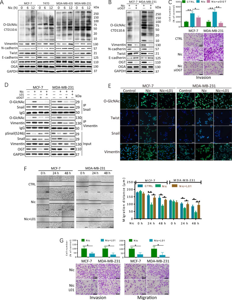
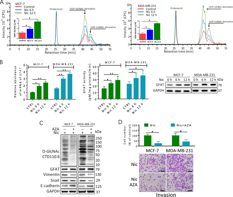
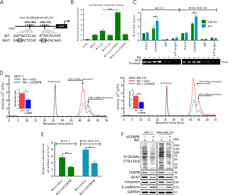
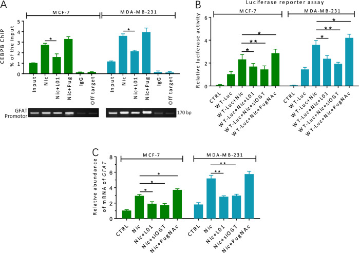
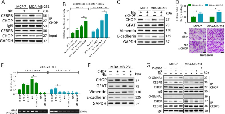
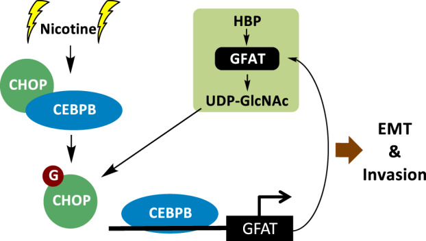
Cigarette smoking has been shown to be a carcinogenic factor in breast cancer. Nicotine (Nic), an active component of tobacco, has been found to induce epithelial-mesenchymal transition (EMT) in breast cancer cells. However, the alterations in protein O-GlcNAcylation in Nic-mediated tumorigenesis and malignization mechanisms are less well studied. Herein, we found that cellular O-GlcNAcylation dramatically increased in human breast cancer cells with EMT activation induced by Nic. Elevated O-GlcNAcylation subsequently promoted Nic-induced EMT activation and increased cell migratory abbility. In addition, we demonstrated that a differentiation factor for the mammary epithelium, CCAAT/enhancer-binding protein B (CEBPB), was involved in Nic-induced hyper-O-GlcNAcylation via transcriptional regulation of the expression of the key enzyme glutamine: fructose-6-phosphate amidotransferase (GFAT) and thus increased the flux through the hexosamine biosynthetic pathway (HBP). Finally, elevated O-GlcNAcylation of the transcriptional repressor C/EBP homologous protein (CHOP) suppressed its heterodimerization with CEBPB and facilitated the DNA-binding activity of CEBPB, further generating positive feedback that enhanced EMT upon Nic stimulation. In conclusion, our results have revealed a new regulatory mechanism involving CEBPB/GFAT-induced hyper-O-GlcNAcylation that plays a key role in EMT and smoking-mediated breast cancer progression.


Conflict of interest statement

The authors declare that they have no conflict of interest.

Figures

Fig. 1
Fig. 1. Hyper-O-GlcNAcylation levels contribute to Nic-induced EMT and invasion in breast cancer cells.
a O-GlcNAcylation was upregulated in breast cancer cells after treatment with Nic. Breast cancer cells were treated with 100 μM Nic for the indicated times. Cellular O-GlcNAcylation and the expression of EMT markers in breast cancer cell lines were analyzed by immunoblotting analysis. (b, c) MCF-7 and MDA-MB-231 cells were transfected with scrambled siRNA (−) or OGT siRNA (siOGT, +) for 48 h and then treated with 100 μM Nic for an additional 24 h. Protein levels were examined by immunoblotting. Transwell invasion assays showed that the reduction of O-GlcNAcylation inhibited the Nic-induced invasion of breast cancer cells. d The EMT-related proteins Snail and Vimentin were O-GlcNAcylated during Nic treatment. MCF-7 and MDA-MB-231 cells were transfected with scrambled siRNA (−) or OGT siRNA (siOGT, +) for 48 h and then treated with 100 μM Nic for 24 h. 100 μM L01 was used to inhibit O-GlcNAcylation. Snail and Vimentin immunoprecipitations assays were performed, and the immunoprecipitated fractions were analyzed by immunoblotting. e Immunocytochemistry of MCF-7 and MDA-MB-231 cells treated with 100 μM Nic either alone or together with 100 μM L01 for 12 h. The immunoreactivity of the antibodies for O-GlcNAc CTD110.6 and EMT markers is shown in green, and DAPI is shown in blue. f Wound healing assays and Transwell migration and invasion assays showed that the reduction of O-GlcNAcylation inhibited the Nic-induced migration and invasion of breast cancer cells. Cells were treated with 100 μM Nic either alone or together with 100 μM L01 for 24 h and 48 h. Graphs comparing the average migration rate in different treated cells are shown. The data represent the mean ± SEM, N = 3, *p < 0.05, **p < 0.01
Fig. 2
Fig. 2. The HBP flux and GFAT are upregulated in breast cancer cells in response to Nic.
a Nic induced increases in UDP-GlcNAc levels. UDP-GlcNAc in the cell lysates was derivatized with trimethylsilyldiazomethane. Chromatograms of polar metabolites in MCF-7 and MDA-MB-231 cell extracts from control cells (red line) and cells treated with 100 μM Nic for 6 h (blue line) or 12 h (green line) show regions corresponding to the UDP-GlcNAc derivative, UDP-GalNAc derivative and probenecid retention times. Quantitative analyses are shown. Quantitative analyses are shown with the standard deviation based on three independent experiments P-values were calculated using one-way ANOVA and the appropriate post test. *P < 0.05. b GFAT transcriptional levels, protein levels and activity were upregulated in breast cancer cells after treatment with Nic for the indicated times. The GFAT transcript level was analyzed by quantitative RT-PCR. DMSO was used as a control (CTRL). Whole-cell lysates were analysed by immunoblotting. Cell lysates were also used for the analysis of GFAT activity. c, d MCF-7 and MDA-MB-231 cells were treated with 100 μM Nic either alone or together with 25 μM AZA for 24 h. Protein levels were examined by immunoblotting. DMSO was used as a control (CTRL). Transwell invasion assays showed that inhibition of GFAT reduced Nic-induced the invasion of breast cancer cells. The data represent the mean ± SEM, N = 3, *p < 0.05, **p < 0.01
Fig. 3
Fig. 3. GFAT accumulation upon Nic is regulated by CEBPB.
a Schematic description of the putative GFAT distal promoter with potential CEBPB-binding sites. A wild-type GFAT promoter (WT) luciferase reporter construct and a mutant (MUT) form lacking the CEBPB-binding site are shown. b 293T cells were transfected with a reporter vector consisting of luciferase cDNA fused to the GFAT promoter. The pGL3 vector and mutational (MUT) GFAT promoter were used as controls. Transfection of CEBPB increased GFAT promoter activity. c MCF-7 and MDA-MB-231 cells were treated with or without 100 μM Nic for 24 h. The lysates were subjected to ChIP. Quantitative PCR amplification was performed using primers specific for the GFAT promoter. d The induction of UDP-GlcNAc by Nic was attenuated by CEBPB inhibition. Chromatograms of polar metabolites in MCF-7 and MDA-MB-231 cell extracts from cells treated with either 100 μM Nic alone (red line) or CEBPB siRNA plus Nic (blue line) for 24 h show regions corresponding to the UDP-GlcNAc derivative, UDP-GalNAc derivative and probenecid retention times. e GFAT transcriptional levels were reduced in breast cancer cells after CEBPB knockdown. The GFAT transcript level was analyzed by quantitative RT-PCR. Scrambled siRNA was used as a control. f MCF-7 and MDA-MB-231 cells were transfected with scrambled siRNA (-) or OGT siRNA (siCEBPB, +) for 48 h and then treated with 100 μM Nic for an additional 24 h. The protein levels were examined by immunoblotting. The data represent the mean ± SEM, N = 3, *p < 0.05, **p < 0.01
Fig. 4
Fig. 4. O-GlcNAcylation is involved in the control of GFAT transcription in Nic-treated breast cancer cells.
a MCF-7 and MDA-MB-231 cells were treated with 100 μM Nic either alone or together with L01 (100 μM)/PugNAc (50 μM) for 24 h. The lysates were subjected to ChIP assays. Quantitative PCR amplification was performed using primers specific for GFAT promoter. b Indicated cells were transfected with a reporter vector consisting of luciferase cDNA fused to GFAT promotor. Transfection of OGT siRNA or inhibition of O-GlcNAc (100 μM L01) decreased GFAT promotor activity. PugNAc (50 μM) increased GFAT promotor activity. c O-GlcNAcylation regulates GFAT transcription in breast cancer cells with Nic treatment (100 μM, 24 h). Transfection of OGT siRNA or inhibition of O-GlcNAc (100 μM L01) decreased GFAT transcription. PugNAc (50 μM) increased GFAT transcription. GFAT transcript level was analyzed by quantitative RT-PCR. Scrambled siRNA was used as a control (CTRL). The data represent the mean ± SEM, N = 3, *p < 0.05, **p < 0.01
Fig. 5
Fig. 5. O-GlcNAcylation of CHOP suppresses its transcriptional inhibition of CEBPB.
a CHOP/CEBPB dimer is reduced in breast cancer cells with Nic treatment (100 μM, 24 h). CHOP co-IP were performed, and immunoprecipitated fractions were analyzed by immunoblotting for the indicated proteins. b Indicated cells were transfected with a reporter vector consisting of luciferase cDNA fused to GFAT promoter and then treated for 24 h. CHOP knockdown with siRNA (siCHOP) increased GFAT promoter activity. c, d MCF-7 and MDA-MB-231 cells were transfected with scrambled siRNA (−) or CHOP siRNA (siCHOP, +) for 48 h and then treated with 100 μM Nic for an additional 24 h. Protein levels were examined by immunoblotting. Transwell invasion assays showed that reduction of O-GlcNAcylation inhibited Nic-induced invasion in breast cancer cells. e, f MDA-MB-231 cells were treated with 100 μM Nic either alone or together with transfection of pcDNA3-CHOP for 24 h. pcDNA3.1 vector was used as a control (CTRL). The lysates were subjected to ChIP assays with indicated antibodies. Quantitative PCR amplification was performed using primers specific for GFAT promoter. The protein levels were examined by immunoblotting. g O-GlcNAc modification of CHOP is an obstacle for transcriptional inhibition of CEBPB. MCF-7 and MDA-MB-231 cells were treated with 100 μM Nic either alone or together with L01 (100 μM)/PugNAc (50 μM) for 24 h. Immunoprecipitation was performed, and immunoprecipitated fractions were analyzed by immunoblotting for the indicated proteins. The data represent the mean±SEM, N = 3, *p < 0.05
Fig. 6
Fig. 6. Proposed model for EMT and invasion signaling based on Nic-induced activation of O-GlcNAcylation.
Nic stimulates CEBPB-dependent transcription of GFAT and then mediates the production of UDP-GlcNAc and hyper-O-GlcNAcylation. Excess O-GlcNAcylation promotes the activation of EMT and breast cancer cell invasion. The O-GlcNAcylation of CHOP also prevents its inhibition of CEBPB, subsequently enhancing GFAT transcription in a positive feedback manner

References

    1. Grando, S. A. Connections of nicotine to cancer. Nat. Rev. Cancer14, 419–429 (2014). - PubMed
    1. Gaudet, M. M. et al. Pooled analysis of active cigarette smoking and invasive breast cancer risk in 14 cohort studies. Int. J. Epidemiol.46, 881–893 (2017). - PMC - PubMed
    1. Boone, S. D. et al. Active and passive cigarette smoking and mortality among Hispanic and non-Hispanic white women diagnosed with invasive breast cancer. Ann. Epidemiol.25, 824–831 (2015). - PMC - PubMed
    1. Reynolds, P. et al. Active smoking, household passive smoking, and breast cancer: evidence from the California Teachers Study. J. Natl Cancer Inst.96, 29–37 (2004). - PubMed
    1. Passarelli, M. N. et al. Cigarette smoking before and after breast cancer diagnosis: mortality from breast cancer and smoking-related diseases. J. Clin. Oncol.34, 1315–1322 (2016). - PMC - PubMed

Publication types

MeSH terms

Substances