. 2019 Apr 24;9(1):6483.
doi: 10.1038/s41598-019-42906-3.

Propidium iodide staining underestimates viability of adherent bacterial cells

Affiliations

Propidium iodide staining underestimates viability of adherent bacterial cells

Merilin Rosenberg et al. Sci Rep. .

Abstract

Combining membrane impermeable DNA-binding stain propidium iodide (PI) with membrane-permeable DNA-binding counterstains is a widely used approach for bacterial viability staining. In this paper we show that PI staining of adherent cells in biofilms may significantly underestimate bacterial viability due to the presence of extracellular nucleic acids (eNA). We demonstrate that gram-positive Staphylococcus epidermidis and gram-negative Escherichia coli 24-hour initial biofilms on glass consist of 76 and 96% PI-positive red cells in situ, respectively, even though 68% the cells of either species in these aggregates are metabolically active. Furthermore, 82% of E. coli and 89% S. epidermidis are cultivable after harvesting. Confocal laser scanning microscopy (CLSM) revealed that this false dead layer of red cells is due to a subpopulation of double-stained cells that have green interiors under red coating layer which hints at eNA being stained outside intact membranes. Therefore, viability staining results of adherent cells should always be validated by an alternative method for estimating viability, preferably by cultivation.


Conflict of interest statement

The authors declare no competing interests.

Figures
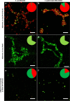
Figure 1
Figure 1
Epifluorescence microscopy images of adherent E. coli (a,c,e)  and S. epidermidis (b,d,f) viability staining. 24 h initial monolayer biofilm formed on glass in PBS stained in situ with propidium iodide (PI) and SYTO 9 (a,b), with fluorescein diacetate (FDA) (c,d) or harvested via sonication, stained with PI and SYTO 9 and collected on filter (e,f). Pie diagrams represent total cell count on surfaces with PI, SYTO 9 and FDA stained signal proportions marked in red, dark green and light green respectively. Scale bars correspond to 10 µm.
Figure 2
Figure 2
Comparison of multiple approaches to evaluate adherent cell viability in E. coli (a,c) and S. epidermidis (b,d) biofilms on surface in situ (a,b) or after harvesting via ultrasonication (c,d). 24 h initial monolayer biofilm formed on glass in PBS stained in situ (a,b) with propidium iodide (PI) and SYTO 9 or FDA followed by epifluorescence microscopy (EM) and signal counting or harvested (c,d) and cultivated for plate counts, co-stained with PI and SYTO 9 and analyzed by flow cytometry (FCM) or collected on filter followed by EM and signal counting. Cell counting results are presented as signals/cm2 where one signal counted corresponds to a single fluorescent cell or compact diplococcus (microscopy), a CFU (cultured plate count) or an FCM event. Live/dead gating of FCM signal populations was based on known proportions of viable and ethanol-killed planktonic bacteria. Mean and standard deviation of 4–6 independent values for in situ staining and filtering and 10–16 independent values for plate counts and FCM are shown and only statistically significant differences (p < 0.05) marked on graphs (“ “ > 0.05; * < 0.05; ** < 0.01; *** < 0.001; **** < 0.0001).
Figure 3
Figure 3
Confocal laser scanning microscopy (CSLM) images of 24 h E. coli biofilm co-stained with propidium iodide (PI) and SYTO 9: vertical and horizontal cross-sections in multichannel (a), green channel (b) and red channel (c) view. Dead cells stained with PI are indicated with cyan and viable cells double-stained with PI and SYTO 9 with yellow arrows. Scale bars correspond to 5 µm. Single images of the Z-stack are available in Supplementary Album 1.
Figure 4
Figure 4
Confocal laser scanning microscopy (CSLM) images of 24 h S. epidermidis biofilm co-stained with propidium iodide (PI) and SYTO 9: vertical and horizontal cross-sections in multichannel (a), green channel (b) and red channel (c) view. Dead cells stained with PI are indicated with cyan and viable cells double-stained with PI and SYTO 9 with yellow arrows. Scale bars correspond to 5 µm. Single images of the Z-stack are available in Supplementary Album 1.

References

    1. Boulos L, Prévost M, Barbeau B, Coallier J, Desjardins R. LIVE/DEAD® BacLightTM: application of a new rapid staining method for direct enumeration of viable and total bacteria in drinking water. J. Microbiol. Methods. 1999;37:77–86. doi: 10.1016/S0167-7012(99)00048-2. - DOI - PubMed
    1. Stocks SM. Mechanism and use of the commercially available viability stain,BacLight. Cytometry. 2004;61A:189–195. doi: 10.1002/cyto.a.20069. - DOI - PubMed
    1. User Manual: LIVE/DEAD BacLight Bacterial Viability Kits. (2004).
    1. Stiefel P, Schmidt-Emrich S, Maniura-Weber K, Ren Q. Critical aspects of using bacterial cell viability assays with the fluorophores SYTO9 and propidium iodide. BMC Microbiol. 2015;15:36. doi: 10.1186/s12866-015-0376-x. - DOI - PMC - PubMed
    1. Kirchhoff C, Cypionka H. Propidium ion enters viable cells with high membrane potential during live-dead staining. J. Microbiol. Methods. 2017;142:79–82. doi: 10.1016/j.mimet.2017.09.011. - DOI - PubMed

Publication types

MeSH terms

LinkOut - more resources