Review
. 2019 Sep;19(9):539-549.
doi: 10.1038/s41577-019-0167-y.

How macrophages deal with death

Affiliations
Review

How macrophages deal with death

Greg Lemke. Nat Rev Immunol. 2019 Sep.

Abstract

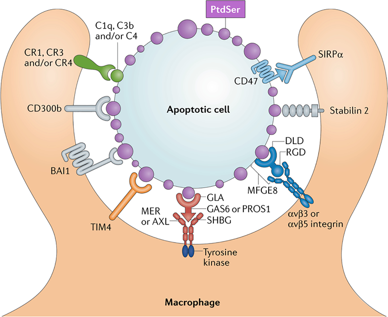
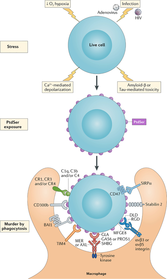
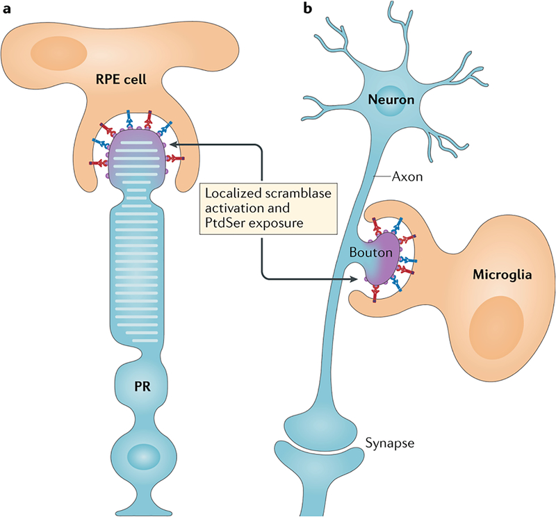
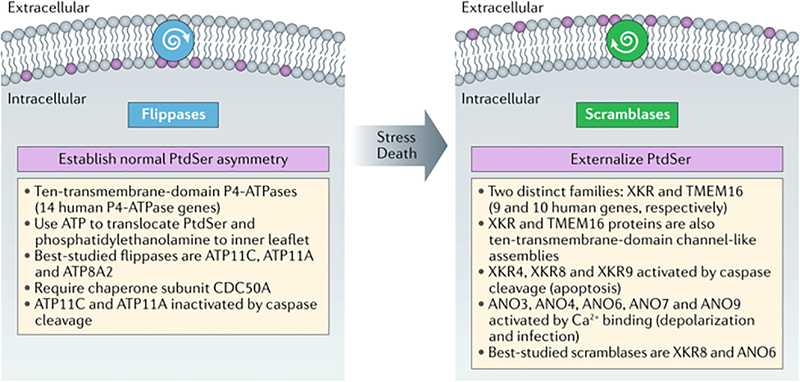
Tissue macrophages rapidly recognize and engulf apoptotic cells. These events require the display of so-called eat-me signals on the apoptotic cell surface, the most fundamental of which is phosphatidylserine (PtdSer). Externalization of this phospholipid is catalysed by scramblase enzymes, several of which are activated by caspase cleavage. PtdSer is detected both by macrophage receptors that bind to this phospholipid directly and by receptors that bind to a soluble bridging protein that is independently bound to PtdSer. Prominent among the latter receptors are the MER and AXL receptor tyrosine kinases. Eat-me signals also trigger macrophages to engulf virus-infected or metabolically traumatized, but still living, cells, and this 'murder by phagocytosis' may be a common phenomenon. Finally, the localized presentation of PtdSer and other eat-me signals on delimited cell surface domains may enable the phagocytic pruning of these 'locally dead' domains by macrophages, most notably by microglia of the central nervous system.


Conflict of interest statement

Competing interests

The author declares no competing interests.

Figures

Figure 1.
Figure 1.. Receptors and ligands mediating apoptotic cell (AC) recognition and phagocytosis by macrophages.
Apoptotic cell surfaces are marked by a profusion of membrane blebs that universally display externalized phosphatidylserine (PtdSer, purple). PtdSer – the most potent ‘eat-me’ signal for apoptotic cell phagocytosis – is recognized by macrophage receptors that directly bind this phospholipid – including CD300b, BAI1, TIM4 (green), and Stabilin-2. PtdSer is also recognized by soluble, bi-functional ‘bridging’ proteins, including Gas6/Pros1 (red) and MFG-E8 (blue). These proteins carry one domain that binds PtdSer – the ‘Gla’ domains of Gas6/Pros1 and the Discoidin-like domain (Dld) of MFG-E8 – and a second domain that binds to phagocytic receptors expressed by macrophages – the SHBG domain of Gas6/Pros1 and an RGD motif within the second EGF-like domain of MFG-E8. The macrophage receptors for Gas6/Pros1 are the TAM receptor tyrosine kinases Mer and Axl (red), while those for MFG-E8 are αvβ3 and αvβ5 integrin dimers (blue). Complement proteins, including C1q, C3b, and C4 (closed gray circle), also decorate the surface of apoptotic cells by mechanisms that remain under study, but may involve the recognition of PtdSer. These complement factors are recognized by the complement receptors CR1, CR3, and CR4. Some apoptotic cells also express CD47 (light blue), which negatively regulates phagocytosis by acting as a ‘don’t-eat-me’ signal. Its receptor is SIRPα. All phagocytic receptors must engage signal transduction networks that ultimately activate Rho family GTPases, but the only receptors that carry intrinsic signaling activity are the TAM receptors, which are robust, ligand/apoptotic cell-activated tyrosine kinases (TKs).
Figure 2.
Figure 2.. PtdSer-triggered phagocytic engulfment of living cells.
Recent findings indicate that a variety of stressors - including hypoxia precipitated by stroke, calcium influx triggered by excitatory and/or exitotoxic stimulation (depolarization), infection by HIV and adenoviruses (AdV), and the toxic effects of Amyloid β (Aβ) and phospho-Tau deposition (top) – can lead to the activation of XKR and TMEM16 scramblases and the exposure of PtdSer on the surface of living cells (middle). In some settings, this leads to the engulfment of stressed but still living cells – or murder by phagocytosis (bottom).
Figure 3.
Figure 3.. PtdSer-delimited phagocytosis of only small parts of cells.
a. The phagocytic retinal pigment epithelial (RPE) cells of the eye pinch of and engulf the distal ends of the rhodopsin-containing outer segments of photoreceptors (PR) each morning. This localized phagocytosis is entirely Mer-dependent (red) and is assisted by the MFG-E8 integrin system (blue). Patients with complete loss-of-function MERTK mutations have an inherited form of retinitis pigmentosa due to a failure in RPE phagocytosis. b. Microglia, the tissue macrophages of the brain, phagocytose unpaired pre-synaptic axonal boutons of the postnatal brain (those that are not part of an anatomically complete, functional synapse), in a process termed ‘synaptic pruning’. These boutons are decorated by several of the eat-me signals highlighted in Fig. 1, including complement proteins (gray). Importantly, the extent of the bouton to be pruned may again be delimited by the local externalization of PtdSer on its surface. The localized activation of scramblase activity for the events depicted in a and b is at this point a speculation.
None

Similar articles

Cited by

References

    1. Nagata S Apoptosis and Clearance of Apoptotic Cells. Annu Rev Immunol 36, 489–517 (2018). - PubMed
    1. Elliott MR & Ravichandran KS The Dynamics of Apoptotic Cell Clearance. Dev Cell 38, 147–160 (2016). - PMC - PubMed
    1. Gordon S & Pluddemann A Macrophage Clearance of Apoptotic Cells: A Critical Assessment. Front Immunol 9, 127 (2018). - PMC - PubMed
    1. Epelman S, Lavine KJ & Randolph GJ Origin and functions of tissue macrophages. Immunity 41, 21–35 (2014). - PMC - PubMed
    1. Krysko DV, Vanden Berghe T, D’Herde K & Vandenabeele P Apoptosis and necrosis: detection, discrimination and phagocytosis. Methods 44, 205–221 (2008). - PubMed

Publication types