. 2019 Apr 4:13:317.
doi: 10.3389/fnins.2019.00317. eCollection 2019.

Evidence of Aquaporin 4 Regulation by Thyroid Hormone During Mouse Brain Development and in Cultured Human Glioblastoma Multiforme Cells

Affiliations

Evidence of Aquaporin 4 Regulation by Thyroid Hormone During Mouse Brain Development and in Cultured Human Glioblastoma Multiforme Cells

Lucas E S Costa et al. Front Neurosci. .

Abstract

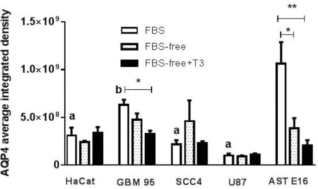
Accumulating evidence indicates that thyroid function and the thyroid hormones L-thyroxine (T4) and L-triiodothyronine (T3) are important factors contributing to the improvement of various pathologies of the central nervous system, including stroke, and various types of cancer, including glioblastoma multiforme (GBM). Low levels of T3 are correlated with the poorest outcome of post-stroke brain function, as well as an increased migration and proliferation of GBM tumor cells. Thyroid hormones are known to stimulate maturation and brain development. Aquaporin 4 (AQP4) is a key factor mediating the cell swelling and edema that occurs during ischemic stroke, and plays a potential role in the migration and proliferation of GBM tumor cells. In this study, as a possible therapeutic target for GBM, we investigated the potential role of T3 in the expression of AQP4 during different stages of mouse brain development. Pregnant mice at gestational day 18, or young animals at postnatal days 27 and 57, received injection of T3 (1 μg/g) or NaOH (0.02 N vehicle). The brains of mice sacrificed on postnatal days 0, 30, and 60 were perfused with 4% paraformaldehyde and sections were prepared for immunohistochemistry of AQP4. AQP4 immunofluorescence was measured in the mouse brains and human GBM cell lines. We found that distribution of AQP4 was localized in astrocytes of the periventricular, subpial, and cerebral parenchyma. Newborn mice treated with T3 showed a significant decrease in AQP4 immunoreactivity followed by an increased expression at P30 and a subsequent stabilization of aquaporin levels in adulthood. All GBM cell lines examined exhibited significantly lower AQP4 expression than cultured astrocytes. T3 treatment significantly downregulated AQP4 in GBM-95 cells but did not influence the rate of GBM cell migration measured 24 h after treatment initiation. Collectively, our results showed that AQP4 expression is developmentally regulated by T3 in astrocytes of the cerebral cortex of newborn and young mice, and is discretely downregulated in GBM cells. These findings indicate that higher concentrations of T3 thyroid hormone would be more suitable for reducing AQP4 in GBM tumorigenic cells, thereby resulting in better outcomes regarding the reduction of brain tumor cell migration and proliferation.

Keywords: GBM; aquaporin 4; brain development; brain tumor; thyroid hormone.


Figures

FIGURE 1
FIGURE 1
(Upper) Aquaporin 4 (AQP4) distribution in the cerebral cortex of mice at postnatal days 0, 30, and 60 in control (CTRL) and T3-injected animals. Insets depict AQP4 immunoreactivity (green: A, C, E, G, I, and K) or merged GFAP (red) colocalization (B, D, F, H, J, and L). Immunoreactivity of AQP4 showed polarization to perivascular astrocytic end feet surrounding the cerebral microvessels (arrowheads in CTRL P0 and T3 P30) or in subpial astrocytes as depicted in CTRL P30 and T3 P0 slices. T3 injection caused an increase in AQP4 immunoreactivity with developmental progression. (Lower) AQP4 fluorescence levels in coronal sections of total brains. Mice treated with T3 showed higher levels of AQP4 from 30 to 60 days of age. N = 3 or 4 for each group; fluorescence analysis from 6 to 10 coronal sections of 40-μm thickness. Student’s t-test; ∗∗P < 0.01; ∗∗∗∗P < 0.0001; #P < 0.0001 one-way ANOVA followed by Tukey’s test (CTRL P0 vs. CTRL P60; T3 P0 vs. T3 P30 and T3 P60).
FIGURE 2
FIGURE 2
Phase-contrast microscopic images of glioblastoma multiforme GBM-11 and GBM-95 cells cultured in medium containing 10% fetal bovine serum (FBS), medium without serum (FBS-free), and treated with T3 in medium without serum (FBS-free+T3). GBM tumor cells treated with T3 showed large cytoplasmic volume (E,F, white arrows) and processes (E,F, black arrows) when compared with those maintained in FBS-free medium (C,D). GBM-11 and GBM-95 cells maintained in serum-containing medium (A,B).
FIGURE 3
FIGURE 3
Phase-contrast microscopic images of HaCat and SCC-4 cells in medium containing 10% FBS, medium without serum (FBS-free), and treated with T3 in medium without serum (FBS-free+T3). In the SCC-4 line, cells grown in T3-treated and FBS-free medium showed more disorganized cell–cell contacts (C,D) compared with those of cells maintained in serum-containing medium (A,B). White arrows indicate large cytoplasmic volume (D,F) and black arrows cytoplasmic processes (C,E).
FIGURE 4
FIGURE 4
Fluorescence analysis for AQP4 in HaCat, GBM-95, SCC-4, and U-87 cell lines and a secondary culture of astrocytes (AST E16). The integrated density is the sum of the values of the pixels in the area considered fluorescent. T3 treatment significantly downregulated AQP4 in GBM-95 cells maintained in FBS. Among all the cell lines examined, astrocytes exhibited the highest expression of AQP4. T3 treatment in FBS-free medium reduced AQP4 expression in astrocytes. HaCat, SCC-4, and U87 cell lines showed no change in AQP4 expression. Cells were cultured in medium containing 10% FBS, medium without serum (FBS-free), and treated with T3 in medium without serum (FBS-free+T3). P < 0.05, ∗∗P < 0.01, ∗∗∗P < 0.001; and (a) P < 0.001; (b) P < 0.05 are related to differences between the tumor cells in FBS medium vs. secondary culture of Ast E16 in FBS.
FIGURE 5
FIGURE 5
(A) Immunofluorescence staining for AQP4 (red) and phalloidin (green) or DAPI (blue). T3 treatment (50 mM) negatively regulated AQP4 expression only in GBM-95 cells when compared to AQP4 expression in cells maintained in FBS, as shown in Figure 6. Cells were cultured in medium containing 10% FBS, medium without serum (FBS-free), and treated with T3 in medium without serum (FBS-free+T3). (B) Immunofluorescence staining for AQP4 (red) and phalloidin (green) or DAPI (blue). HaCat, SCC-4, and U87 cell lines treated with T3 (50 mM) showed no change in AQP4 expression.
FIGURE 6
FIGURE 6
(Upper) Analysis of cell migration in GBM-95 cells and cultured E16 astrocyte cells. The migration and invasion of the GBM cell line was not significantly altered by treatment with T3, whereas the migration of cultured astrocytes was significantly decreased by 24.47% in response to treatment with T3. (Lower) Cell migration assay of GBM-95 cells and cultured E16 astrocytes treated with T3. Assay measurements were performed at two different time points: T0, represents the beginning of the treatment and T24 represents 24 h after the start of treatment. Cells were cultured in medium containing 10% FBS, medium without serum (FBS-free), and treated with T3 in medium without serum (FBS-free+T3). The images were analyzed by measuring the reduction in scratch area (∗∗∗P < 0.001: one-way ANOVA).

Similar articles

Cited by

References

    1. Bárez-López S., Guadaño-Ferraz A. (2017). Thyroid hormone availability and action during brain development in rodents. Front. Cell. Neurosci. 11:240 10.3389/fncel.2017.00240 - DOI - PMC - PubMed
    1. Bárez-López S., Obregon M. J., Bernal J., Guadaño-Ferraz A. (2018). Thyroid hormone economy in the perinatal mouse brain: implications for cerebral cortex development. Cereb. Cortex 28 1783–1793. 10.1093/cercor/bhx088 - DOI - PubMed
    1. Bergh J. J., Lin H. Y., Lansing L., Mohamed S. N., Davis F. B., Mousa S., et al. (2005). Integrin alphaVbeta3 contains a cell surface receptor site for thyroid hormone that is linked to activation of mitogen-activated protein kinase and induction of angiogenesis. Endocrinology 146 2864–2871. 10.1210/en.2005-0102 - DOI - PubMed
    1. Calamita G., Moreno M., Ferri D., Silvestri E., Roberti P., Schiavo L., et al. (2007). Triiodothyronine modulates the expression of aquaporin-8 in rat liver mitochondria. J. Endocrinol. 192 111–120. 10.1677/JOE-06-0058 - DOI - PubMed
    1. Davis F. B., Tang H. Y., Shih A., Keating T., Lansing L., Hercbergs A., et al. (2006). Acting via a cell surface receptor, thyroid hormone is a growth factor for glioma cells. Cancer Res. 66 7270–7275. 10.1158/0008-5472.CAN-05-4365 - DOI - PubMed

LinkOut - more resources