

Aminaphtone Efficacy in Primary and Secondary Raynaud's Phenomenon: A Feasibility Study
- PMID: 31019461
- PMCID: PMC6458253
- DOI: 10.3389/fphar.2019.00293
Aminaphtone Efficacy in Primary and Secondary Raynaud's Phenomenon: A Feasibility Study
Abstract
Objectives: The aim of this six-month open feasibility study was to evaluate skin blood perfusion and clinical symptom changes during aminaphtone treatment in patients with either primary or secondary Raynaud's phenomenon to systemic sclerosis.
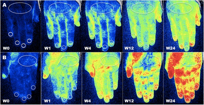
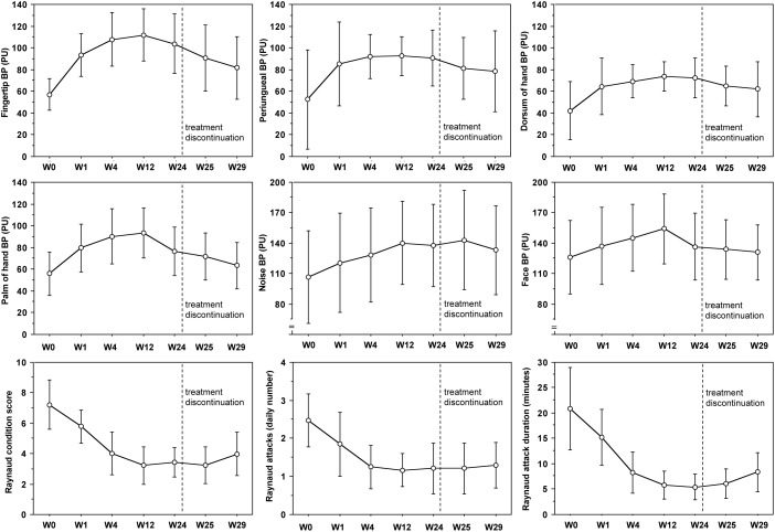
Methods: Ninety-two patients referring for Raynaud's phenomenon have been enrolled in November during routine clinical assessment, after informed consent. Aminaphtone was administered 75 mg twice daily in addition to current treatments to forty-six patients. Skin blood perfusion was measured by Laser Speckle Contrast Analysis (LASCA) at the level of fingertips, periungual areas, dorsum and palm of hands, and face at baseline (W0), after one (W1), four (W4), twelve (W12) and twenty-four (W24) weeks of treatment. Raynaud's condition score (RCS) and both frequency and duration of Raynaud's attacks were assessed at the same time.
Results: Compared with the control group, despite colder period of the year, aminaphtone treated patients showed a progressive statistically significant increase of blood perfusion, as well as a decrease of RCS, frequency of Raynaud's attacks/day and their duration, from W0 to W12 in all skin areas. From W12 to W24 no further increase of blood perfusion was observed. The results were similar in both primary and secondary Raynaud's phenomenon patients. Five weeks after aminaphtone discontinuation blood perfusion values were significantly higher than those at baseline in the majority of skin areas.
Conclusion: This study demonstrates that aminaphtone treatment increases skin blood perfusion and improves Raynaud's phenomenon clinical symptoms, with sustained efficacy up to 6 months, even in patients with systemic sclerosis. A randomized, blind, controlled, clinical trial including a larger number of subjects is advisable to confirm these early results.
Keywords: Raynaud condition score; Raynaud phenomenon; aminaphtone; blood perfusion; clinical symptoms; laser speckle contrast analysis; microcirculation; systemic sclerosis.
Figures
References
-
- Abou-Raya A., Abou-Raya S., Helmii M. (2008). Statins: potentially useful in therapy of systemic sclerosis-related Raynaud’s phenomenon and digital ulcers. J. Rheumatol. 35 1801–1808. - PubMed
-
- Bose N., Bena J., Chatterjee S. (2015). Evaluation of the effect of ambrisentan on digital microvascular flow in patients with systemic sclerosis using laser Doppler perfusion imaging: a 12-week randomized double-blind placebo controlled trial. Arthritis Res. Ther. 17:44. 10.1186/s13075-015-0558-9 - DOI - PMC - PubMed
LinkOut - more resources
Full Text Sources