

Recent developments in advanced imaging in gout
- PMID: 31019573
- PMCID: PMC6469273
- DOI: 10.1177/1759720X19844429
Recent developments in advanced imaging in gout
Abstract
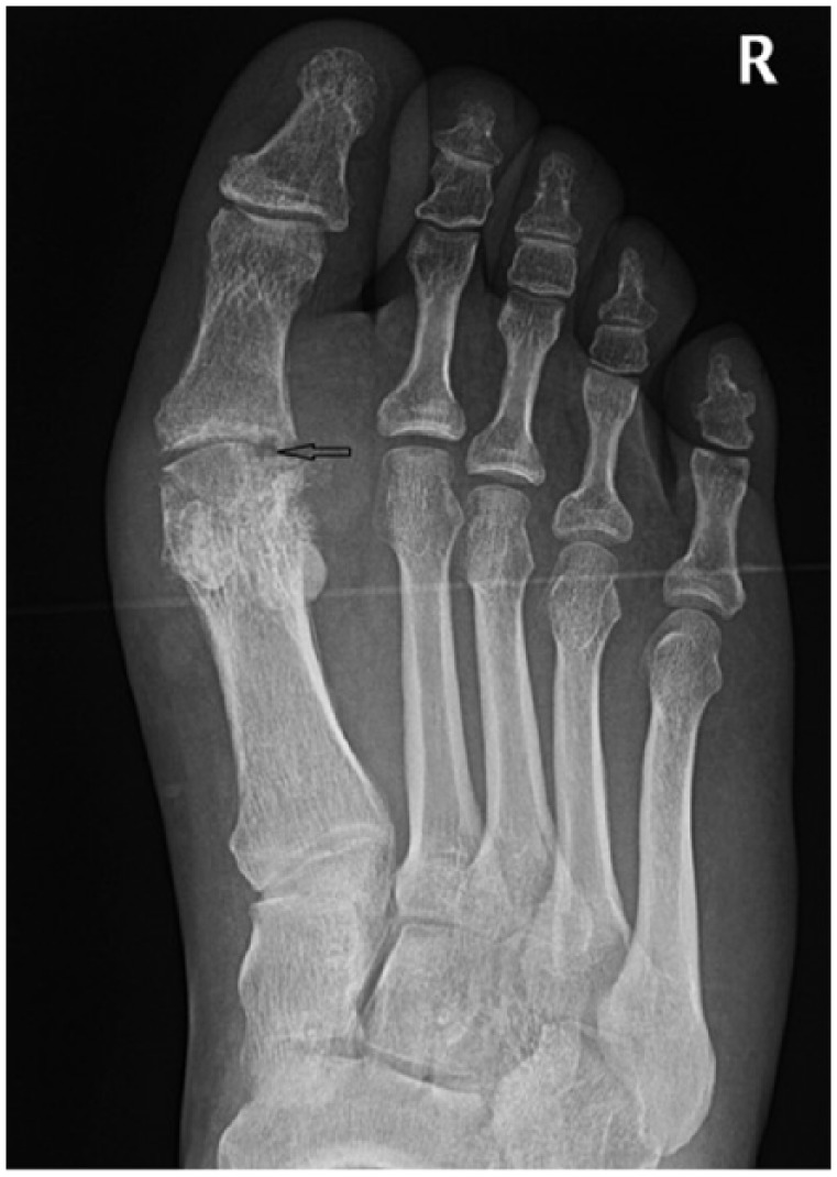
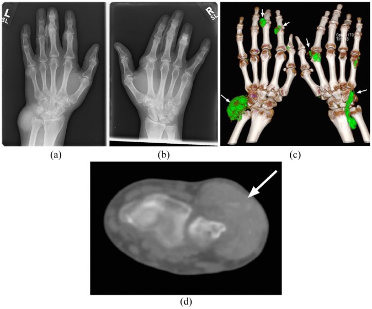
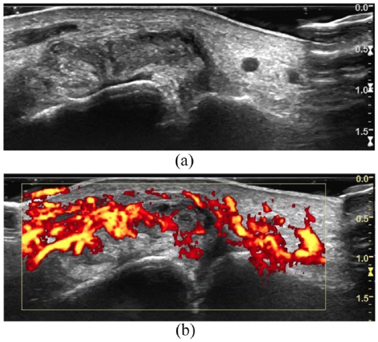
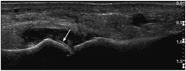
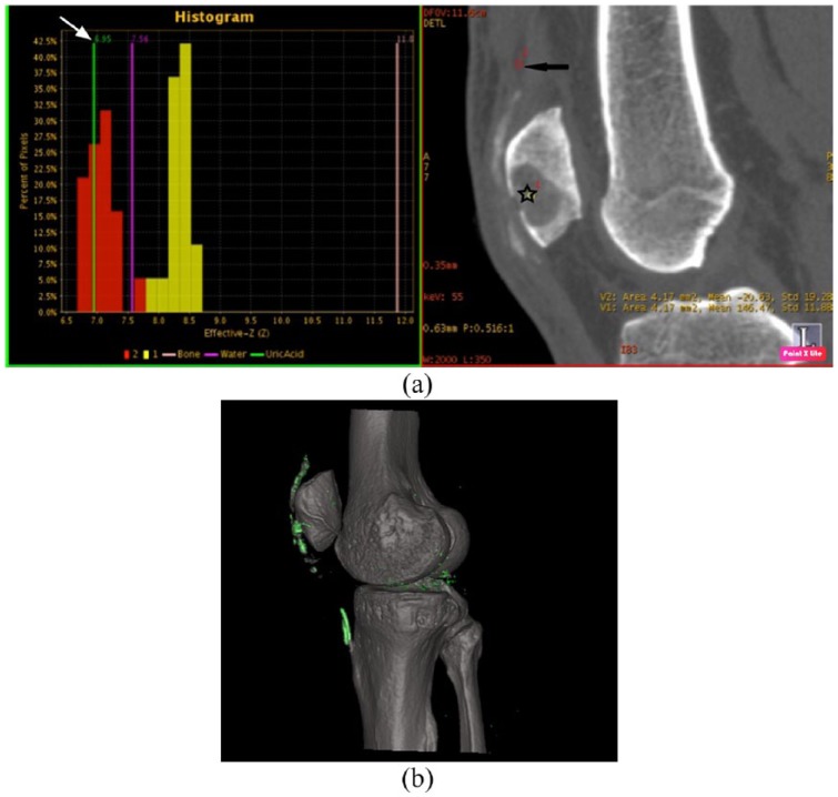
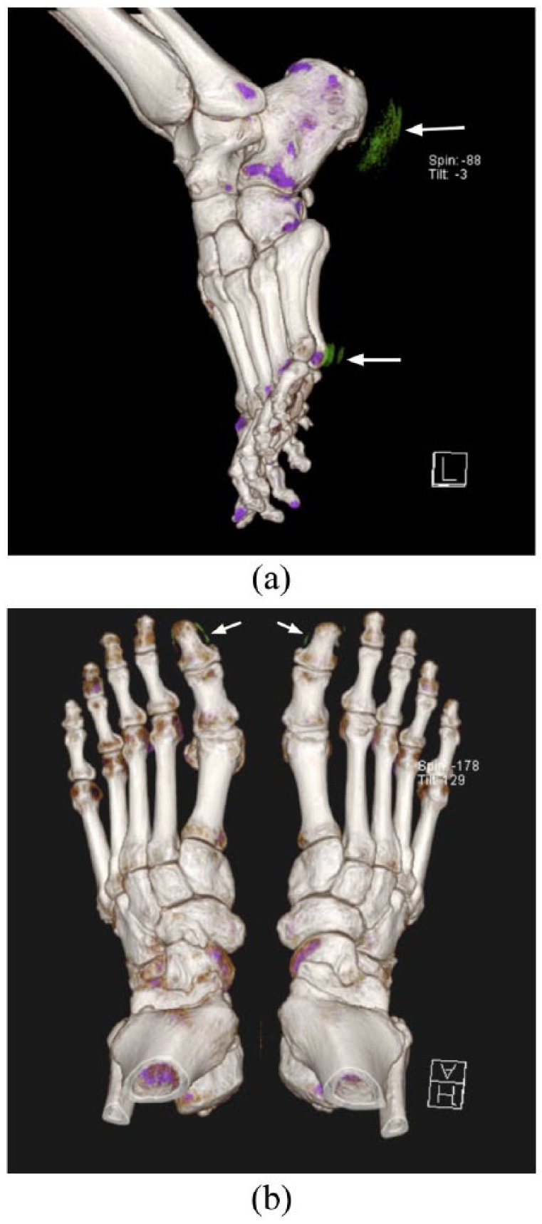
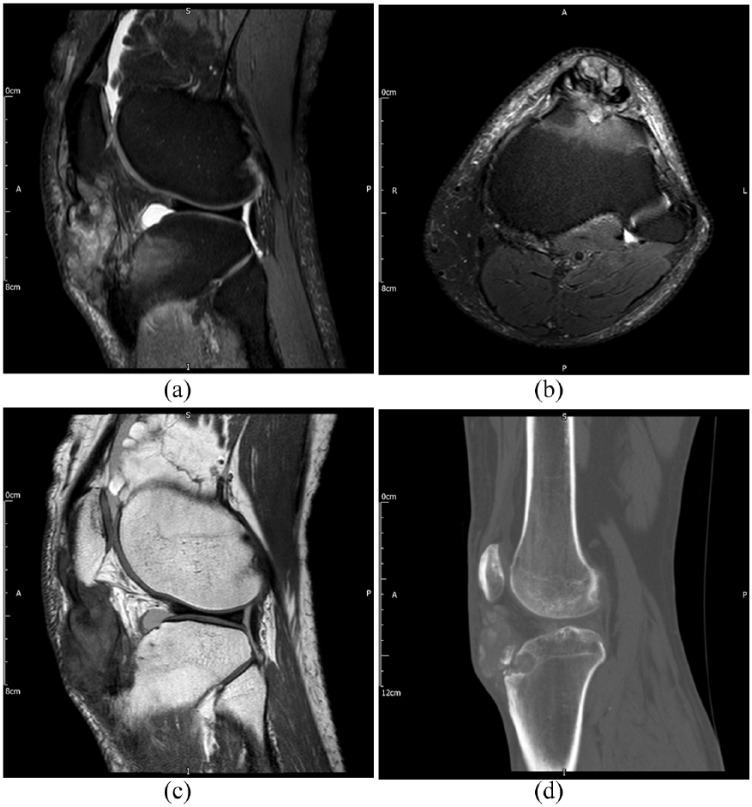
The plain radiographic features of gout are well known; however, the sensitivity of plain radiographs alone for the detection of signs of gout is poor in acute disease. Radiographic abnormalities do not manifest until late in the disease process, after significant joint and soft tissue damage has already occurred. The advent of dual-energy computed tomography (DECT) has enabled the non-invasive diagnosis and quantification of gout by accurately confirming the presence and extent of urate crystals in joints and soft tissues, without the need for painful and often unreliable soft tissue biopsy or joint aspiration. Specific ultrasound findings have been identified and may also be used to aid diagnosis. Both ultrasound and magnetic resonance imaging (MRI) may be used for the measurement of disease extent, monitoring of disease activity or treatment response, although MRI findings are nonspecific. In this article we summarize the imaging findings and diagnostic utility of plain radiographs, ultrasound, DECT, MRI and nuclear medicine studies in the assessment as well as the implications and utility these tools have for measuring disease burden and therapeutic response.
Keywords: crystal arthropathy; diagnostic imaging; dual-energy computed tomography; gout; magnetic resonance imaging; ultrasound.
Conflict of interest statement
Conflict of interest statement: The author(s) declared no potential conflicts of interest with respect to the research, authorship, and/or publication of this article.
Figures
Similar articles
-
Imaging in gout: A review of the recent developments.Ther Adv Musculoskelet Dis. 2014 Aug;6(4):131-43. doi: 10.1177/1759720X14542960. Ther Adv Musculoskelet Dis. 2014. PMID: 25342993 Free PMC article. Review.
-
Gouty arthropathy: Review of clinico-pathologic and imaging features.J Med Imaging Radiat Oncol. 2016 Feb;60(1):9-20. doi: 10.1111/1754-9485.12356. Epub 2015 Oct 5. J Med Imaging Radiat Oncol. 2016. PMID: 26439321 Review.
-
Imaging of gout: New tools and biomarkers?Best Pract Res Clin Rheumatol. 2016 Aug;30(4):638-652. doi: 10.1016/j.berh.2016.10.010. Epub 2016 Nov 12. Best Pract Res Clin Rheumatol. 2016. PMID: 27931959 Review.
-
Tophaceous Gout in an Anorectic Patient Visualized by Dual Energy Computed Tomography (DECT).Am J Case Rep. 2016 Jul 15;17:494-8. doi: 10.12659/ajcr.898542. Am J Case Rep. 2016. PMID: 27418121 Free PMC article.
-
Use of imaging to evaluate gout and other crystal deposition disorders.Curr Opin Rheumatol. 2009 Mar;21(2):124-31. doi: 10.1097/BOR.0b013e3283257b6c. Curr Opin Rheumatol. 2009. PMID: 19339922 Review.
Cited by
-
Single photon emission computed tomography/computed tomography imaging of gouty arthritis: A new voice.J Transl Int Med. 2023 Mar 19;12(3):215-224. doi: 10.2478/jtim-2022-0066. eCollection 2024 Jun. J Transl Int Med. 2023. PMID: 39081275 Free PMC article.
-
MRI of diffuse-type tenosynovial giant cell tumour in the knee: a guide for diagnosis and treatment response assessment.Insights Imaging. 2023 Feb 1;14(1):22. doi: 10.1186/s13244-023-01367-z. Insights Imaging. 2023. PMID: 36725759 Free PMC article. Review.
-
High-Resolution Peripheral Quantitative Computed Tomography for Bone Evaluation in Inflammatory Rheumatic Disease.Front Med (Lausanne). 2020 Jul 15;7:337. doi: 10.3389/fmed.2020.00337. eCollection 2020. Front Med (Lausanne). 2020. PMID: 32766262 Free PMC article.
-
A rare case of gouty arthropathy in the spine complicated with acute cord compression.BJR Case Rep. 2022 May 13;8(4):20220048. doi: 10.1259/bjrcr.20220048. eCollection 2022 Jul 1. BJR Case Rep. 2022. PMID: 36451904 Free PMC article.
-
Limited Knee-Joint Range of Motion in Patients With Tophaceous Gout Improved With Medical Treatment: A 18-Months Follow Up.Front Med (Lausanne). 2020 Feb 28;7:74. doi: 10.3389/fmed.2020.00074. eCollection 2020. Front Med (Lausanne). 2020. PMID: 32181257 Free PMC article.
References
-
- Kuo C-F, Grainge MJ, Zhang W, et al. Global epidemiology of gout: prevalence, incidence and risk factors. Nat Rev Rheumatol 2015; 11: 649–662. - PubMed
-
- Zhu Y, Pandya BJ, Choi HK. Prevalence of gout and hyperuricemia in the US general population: The National Health and Nutrition Examination Survey 2007–2008. Arthritis Rheum 2011; 63: 3136–3141. - PubMed
-
- Beyl RN, Hughes L, Morgan S. Update on importance of diet in gout. Am J Med 2016; 129: 1153–1158. - PubMed
-
- Major TJ, Dalbeth N, Stahl EA, et al. An update on the genetics of hyperuricaemia and gout. Nat Rev Rheumatol 2018; 14: 341–353. - PubMed
Publication types
LinkOut - more resources
Full Text Sources
