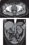
Imaging findings of congenital anomalies of seminal vesicles
- PMID: 31019591
- PMCID: PMC6479056
- DOI: 10.5114/pjr.2019.82711
Imaging findings of congenital anomalies of seminal vesicles
Abstract
The seminal vesicles are paired organs of the male reproductive tract, which produce and secrete seminal fluid. Although congenital anomalies of seminal vesicles are usually asymptomatic, they may lead to various urogenital symptoms, including infertility. Due to their embryologic relationship with other urogenital organs, congenital anomalies of seminal vesicles may accompany other urinary or genital anomalies. Congenital anomalies of seminal vesicles include agenesis, hypoplasia, duplication, fusion, and cyst. These anomalies can be diagnosed with various imaging techniques. The main purpose of this article is to summarise imaging findings and clinical importance of congenital anomalies of seminal vesicles with images of some rare and previously unreported anomalies.
Keywords: computed tomography (CT); congenital anomalies; magnetic resonance imaging; seminal vesicles; ultrasound (US).
Conflict of interest statement
The authors report no conflict of interest.
Figures


Similar articles
-
Magnetic resonance imaging of seminal vesicle cyst associated with ipsilateral urinary anomalies.J Formos Med Assoc. 2006 Feb;105(2):125-31. doi: 10.1016/S0929-6646(09)60333-8. J Formos Med Assoc. 2006. PMID: 16477332
-
The first magnetic resonance imaging-detected case of bilateral seminal vesicle duplication: An illustrative case report.Clin Imaging. 2021 Jun;74:1-3. doi: 10.1016/j.clinimag.2020.12.026. Epub 2021 Jan 4. Clin Imaging. 2021. PMID: 33421696
-
CT and MRI of congenital anomalies of the seminal vesicles.AJR Am J Roentgenol. 2007 Jul;189(1):130-5. doi: 10.2214/AJR.06.1345. AJR Am J Roentgenol. 2007. PMID: 17579162 Review.
-
Seminal vesicles in focus: An illustrated overview.Curr Probl Diagn Radiol. 2024 Sep-Oct;53(5):624-640. doi: 10.1067/j.cpradiol.2024.04.006. Epub 2024 Apr 21. Curr Probl Diagn Radiol. 2024. PMID: 38692935 Review.
-
Multicystic seminal vesicle with ipsilateral renal agenesis: two cases of Zinner syndrome.Scand J Urol. 2017 Feb;51(1):81-84. doi: 10.1080/21681805.2016.1257650. Epub 2016 Dec 1. Scand J Urol. 2017. PMID: 27905212
Cited by
-
Zinner Syndrome Presenting With Chronic Pelvic Pain and Ejaculatory Dysfunction.IJU Case Rep. 2025 Apr 10;8(4):322-325. doi: 10.1002/iju5.70029. eCollection 2025 Jul. IJU Case Rep. 2025. PMID: 40607473 Free PMC article.
-
Curcumin administration alleviates seminal vesicle damage in type 1 diabetic rats by promoting AQP8 expression through AR activation.Sci Rep. 2025 Jan 30;15(1):3804. doi: 10.1038/s41598-024-74750-5. Sci Rep. 2025. PMID: 39885225 Free PMC article.
-
Zinner syndrome: unilateral renal agenesis with unique findings in a 19-year-old patient from Pakistan - a case report with literature review.Ann Med Surg (Lond). 2025 May 20;87(7):4608-4613. doi: 10.1097/MS9.0000000000003413. eCollection 2025 Jul. Ann Med Surg (Lond). 2025. PMID: 40851999 Free PMC article.
-
A rare presentation of Zinner syndrome as recurrent epididymitis: A case report.SAGE Open Med Case Rep. 2023 Sep 12;11:2050313X231200111. doi: 10.1177/2050313X231200111. eCollection 2023. SAGE Open Med Case Rep. 2023. PMID: 37711962 Free PMC article.
-
Magnetic Resonance Imaging Manifestations in 13 Cases of Seminal Vesicle Tuberculosis.Infect Drug Resist. 2023 Oct 26;16:6871-6879. doi: 10.2147/IDR.S427561. eCollection 2023. Infect Drug Resist. 2023. PMID: 37908784 Free PMC article.
References
-
- Vohra S, Morgentaler A. Congenital anomalies of the vas deferens, epididymis, and seminal vesicles. Urology. 1997;49:313–321. - PubMed
-
- Pace G, Galatioto GP, Guala L, et al. Ejaculatory duct obstruction caused by a right giant seminal vesicle with an ipsilateral upper urinary tract agenesia: an embryologic malformation. Fertil Steril. 2008;89:390–394. - PubMed
-
- Mendez-Probst CE, Pautler SE. Fusion of the seminal vesicles discovered at the time of robot-assisted laparoscopic radical prostatectomy. J Robot Surg. 2010;4:45–47. - PubMed
Publication types
LinkOut - more resources
Full Text Sources
