Review
. 2019 Feb 8:84:e92-e111.
doi: 10.5114/pjr.2019.83101. eCollection 2019.

A systematic approach in the diagnosis of paediatric skull lesions: what radiologists need to know

Affiliations
Review

A systematic approach in the diagnosis of paediatric skull lesions: what radiologists need to know

Gagandeep Choudhary et al. Pol J Radiol. .

Abstract

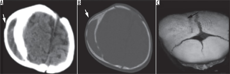
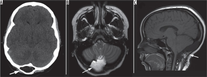
Paediatric skull lesions are commonly identified on imaging. They can be challenging to image, given their location and size, and often require several imaging modalities to narrow down the differential diagnosis. Accurate diagnosis of these lesions is paramount because the clinical therapy can vary tremendously. In this review, we provide a simple and systematic approach to clinical-radiological features of primary skull lesions. We highlight the imaging characteristics and differentiate pathologies based on imaging appearances. We also accentuate the role of cross-sectional imaging in lesion identification and management implications.

Keywords: computed tomography; cross-sectional; magnetic resonance imaging; paediatric skull; skull lesions.


Conflict of interest statement

The authors declare no conflict of interest.

Figures

Figure 1
Figure 1
Imaging pathways of skull lesions in the paediatric population
Figure 2
Figure 2
A) Skull film shows focal depression of right anterior parietal skull (arrow). B, C) Axial head computed tomography with soft tissue and bone windows show abnormal contour and depression of the right calvarium. No acute fractures were identified
Figure 3
Figure 3
Four-month-old girl with frontonasal dysplasia. A, B) Axial head computed tomography images in brain and bone windows show midline nasal and frontal calvarial defects with associated telecanthus. C) 3D reformat and (D) sagittal T1-W images show a large defect in the frontal calvarium
Figure 4
Figure 4
A) Coronal, B) axial head computed tomography angiography show a large left paramidline skull defect (white arrow) and an associated scalp defect, where the superior sagittal sinus abuts and extends to the cutaneous surface (black arrow). C) 3D reformat shows the skull defect and scattered small parietal foramina. D) Clinical image of scalp ulcer/wound
Figure 5
Figure 5
A, B) Sagittal and (C, D) axial head computed tomography images on brain and bone window show a prominent arachnoid granulation with scalloping of the calvarial inner table
Figure 6
Figure 6
Five-year-old girl with multiple arachnoid cysts and clinically visible swelling. A, B) Axial head computed tomography images show extra-axial cerebrospinal fluid (CSF) signal lesion in the left temporal region with scalloping of the inner table of the skull and bulging overlying skull. C, D) Axial T2-W images show extra-axial CSF signal cystic lesions exerting mild mass effect on the adjacent brain parenchyma and skull
Figure 7
Figure 7
One-year-old male patient with epidermoid cyst. A) Frontal X-ray of the skull shows a well-demarcated lytic lesion with sclerotic borders involving the frontal bone at the midline. B-D) Axial T2, axial T1 post contrast, and sagittal T1 images show an oval shaped non-enhancing cystic lesion involving the frontal bone with expansion of the diploic space. E) Ultrasonography shows an oval shaped homogeneous lesion with hypoechogenic internal structure, hyperechogenic surrounding wall, and no obvious flow on colour Doppler
Figure 8
Figure 8
Right periorbital dermoid cyst. A) Frontal X-ray of the skull shows an ill-defined lytic lesion involving the right temporal bone. B-E) Axial T1, T1 post contrast, T2 and T1 fat-sat post-contrast images, respectively, shows a fat-containing lesion expanding the right temporal bone with thin peripheral enhancement
Figure 9
Figure 9
Ten-year-old boy with right parietal foramen. A) Axial computed tomography, B) T2-W, and (C) coronal Gd-enhanced T1-W show a prominent right parietal foramen (arrows)
Figure 10
Figure 10
Occipital encephalocele. A) Sagittal T1-W and (B) axial T2-W images show a small midline defect with a herniated sac containing cerebrospinal fluid and a small portion of the cerebellar parenchyma (arrow). Naso-ethmoidal encephalocele. C) Axial non-enhanced computed tomography and (D) sagittal T1-W images show midline brain tissue mass protruding through a defect in the cribriform plate as well as agenesis of the corpus callosum
Figure 11
Figure 11
Atretic parietal encephalocele. A-C) Sagittal T1-W, post Gd T1W, T2W, and (D) axial T2W images demonstrate a slightly off-midline hyperintense T2 herniation sac through a parietal defect (arrows) and a persistent embryonic falcine sinus (arrow head) directed at the atretic cephalocele. Also note the absence of a straight sinus. E, F) Colour Doppler US images demonstrate the herniated sac containing fibro-fatty tissue
Figure 12
Figure 12
Five-month-old boy with Crouzon’s syndrome and skull abnormality. A, B) Axial head computed tomography images on bone window show diffuse morphological abnormality of the inner table of the skull with areas of discontinuation in the calvarium. C) 3D reformat shows the gross bony abnormality and foramina. VP shunt in place for hydrocephalus decompression
Figure 13
Figure 13
Twelve-year-old girl with neurofibromatosis type I and sphenoid wing dysplasia. A) Axial T2-W and (B) T1-W images show absence of the right sphenoid wing, expanded middle cranial fossa, and displaced contents with resultant right proptosis. C) Sagittal T1-WI shows absence of sella and sphenoid wing
Figure 14
Figure 14
Newborn with both cephalohematoma and subgaleal fluid collection. A, B) Coronal T1- and flair-weighted images shows the presence of a cephalohematoma and subgaleal fluid collection respectively (arrows)
Figure 15
Figure 15
Five-month-old boy with enlarging right skull “mass”. A, B) Axial head computed tomography in soft tissue and bone windows, respectively, shows a peripherally ossified subperiosteal haematoma. C) 3D surface rendered image demonstrates a large ossified lump in the area of concern
Figure 16
Figure 16
A) Two-month-old baby boy with MRSA osteomyelitis of skull and scalp abscess. Left: Axial head computed tomography shows a mixed hyperdense lesion overlying and expanding the right lambdoid suture. Right: Post Gd T1-W image shows enhancing soft tissue with intracranial extension and dural thickening/enhancement (black arrow). B) 16-year-old male with acute sinusitis and Pott’s puffy tumour. Left: axial T2-W and right: Gd-enhanced T1-W images demonstrate peripherally enhancing subgaleal abscess collections with soft tissue swelling and rim-enhancing bifrontal epidural abscesses (white arrow) with associated dural thickening and enhancement
Figure 17
Figure 17
Thirteen-year-old male patient with osteoma of the right occipital bone. A) Lateral projection X-ray of the skull shows a well-defined focal bulging mass at the level of the occipital bone (white arrow) seen as an ovoid dense sclerotic lesion related to the outer table of the right occipital bone on the axial head computed tomography images (B-D)
Figure 18
Figure 18
A, B) Axial non-contrast computed tomography of the head in the soft tissue and bone window shows the presence of a small homogenous lesion of fat density overlying the left occipital bone
Figure 19
Figure 19
A) Axial T2-W, B) Post-Gd T1-W, and (C) sagittal fat-suppressed T2 images show a right occipital subgaleal mass (arrows) with isointense T2 signal and no contrast enhancement. Pathological proven scalp myofibroma
Figure 20
Figure 20
Eighteen-year-old male patient with intraosseous haemangioma. A) Axial head computed tomography shows a well-defined osteolytic lesion with a characteristic sunburst pattern at the vertex. B-D) Axial T2 and axial and sagittal T1 gadolinium enhanced images shows predominantly high T2 signal intensity lesion showing diffuse homogenous enhancement at the level of the left parietal bone
Figure 21
Figure 21
Eleven-month-old boy with infantile haemangioma. A) Sagittal T1-W, B) axial T2-W, post-Gd (C) axial, and (D) coronal T1-W images show a T2 hyperintense and avidly enhancing lesion arising from the frontal calvarium with central flow voids
Figure 22
Figure 22
Seven-year-old boy with Langerhans cell histiocytosis. A) Lateral skull plain radiograph shows a single sharply marginated (bevelled edges) lytic calvarial lesion. Subsequent magnetic resonance imaging. B, C) Pre- and post-Gd T1-WIs show a heterogeneously enhancing intradiploic calvarial mass extending across the inner and outer tables
Figure 23
Figure 23
Eighteen-year-old male with right parietal osteosarcoma. A) Contrast-enhanced head computed tomography shows a right parietal enhancing mass. B) Axial T1-W and (C, D) post-Gd T1-W images show an intensely enhancing extra-axial mass with extracranial (white arrows) and intracranial, extra-axial components. Central non-enhancing area is consistent with necrosis
Figure 24
Figure 24
Three-month-old girl with right sinonasal embryonal rhabdomyosarcoma. A-C) Coronal and axial T1 fat-sat post-contrast and axial T2 fat sat images of the face, respectively, shows a heterogeneously enhancing mass centred at the level of the right maxillary sinus extending to the nasal cavity and masticator space with bone destruction as demonstrated on axial computed tomography post-contrast image of the face (D)
Figure 25
Figure 25
Five-year-old girl with stage IV metastatic neuroblastoma to the calvarium. A) Axial contrast-enhanced computed tomography (CT) of the abdomen shows a bulky and partially-calcified right adrenal neuroblastoma. B) Axial head CT on bone window shows a destructive lesion within the diploic space in the left frontal calvarium with soft tissue components (arrow). C) Axial T2-W image shows corresponding full thickness skull defect and hyperintense signal
Figure 26
Figure 26
Calvarial metastasis in a 15-year-old boy with history of Ewing’s sarcoma of the left distal ulna. A) Sagittal STIR of the left forearm shows the heterogeneously hyperintense primary tumour of distal ulna with associated soft tissue involvement. B, C) Pre- and post-Gd T1-WI demonstrate a destructive calvarial metastasis with heterogeneously iso/hypointense FLAIR signal and avid gadolinium enhancement. D) Sagittal Gd-enhanced T1-WI shows avid enhancement. E, F) Axial DWI and ADC map shows mild true diffusion restriction
Figure 27
Figure 27
Seventeen-year-old boy with multiple venous lakes. A) Axial T2-W show multiple hyperintense lesions (arrows) involving the diploic spaces of the calvarium. B, C) Pre- and post-Gd-enhanced T1-W images show peripheral delayed enhancement
Figure 28
Figure 28
Ten-year-old boy with large left parieto-occipital venous malformation with intracranial developmental venous anomalies. A) Axial T2-W and (B-D) post-Gd T1-W images show mixed T2 high signal lesion with corresponding intense contrast enhancement along left parietal calvarium (arrows in A and B) extending intracranially at occipital and suboccipital regions (arrowhead). Also noted is an associated complex intracranial developmental venous malformation with enlarged veins (arrow in C)
Figure 29
Figure 29
Fifteen-year-old girl with Chiari malformation, status post suboccipital craniectomy for decompression and a post-op intraosseous pseudomeningocele. A) Axial head computed tomography shows an expansile fluid density intraosseous lesion separating the inner and outer table of the occipital calvarium. B) Axial T2-WI shows a suboccipital pseudomeningocele with cerebrospinal fluid signal and intraosseous extension. C) Coronal T1-W image shows changes of suboccipital craniectomy and pseudomeningocele (arrows)
Figure 30
Figure 30
Companion case in an adult patient with history of prior trauma. A, B) Axial computed tomography (CT) images on bone and parenchyma windows show focal encephalomalacia of right frontoparietal lobe with cerebrospinal fluid (CSF) extending and expanding the right parietal bone fracture. Note the scalloping of the fracture margins. C) Coronal CT image shows the extent of encephalomalacia and extension of CSF through the skull defect

Similar articles

Cited by

References

    1. Dupuis O, Silveira R, Dupont C, et al. Comparison of “instrument associated” and “spontaneous” obstetric depressed skull fractures in a cohort of 68 neonates. Am J Obstet Gyencol. 2005;192:165–170. - PubMed
    1. Guha-Ray DK. Intrauterine spontaneous depression of fetal skull: a case report and review of literature. J Reprod Med. 1976;16:321–324. - PubMed
    1. Tavares LA, Kottamasu SR, Ezhuthacha SG, et al. Neonatal skull depression: review of four cases. J Perinatol. 1989;9:423–426. - PubMed
    1. Tibbs RE, Lee DC, Parent AD. Depressed skull fracture. In: Batjer HH, Loftus CM, editors. Textbook of Neurological Surgery: Principles and Practice. Philadelphia: Lippincott Williams and Wilkins; 2003. pp. 1082–1095.
    1. Sunil K, Agrawal DM, Praveen Kumar DM, et al. Congenital depression of the skull in neonate: a case of successful conservative management. J Child Neurol. 2010;25:387–389. - PubMed