

Applicability of color-coded computed tomography images in lung volume reduction surgery planning
- PMID: 31019764
- PMCID: PMC6462717
- DOI: 10.21037/jtd.2019.02.36
Applicability of color-coded computed tomography images in lung volume reduction surgery planning
Abstract
Background: Adequate patient selection is the key to successful lung volume reduction in patients with pulmonary emphysema. Computed tomography (CT) enables a reliable detection of pulmonary emphysema and allows an accurate quantification of the severity. Our goal was to investigate the usefulness and reliability of color-coded (CC) CT images in classification of emphysema and preoperative lung volume reduction planning.
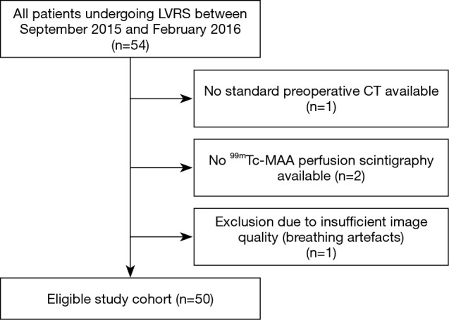
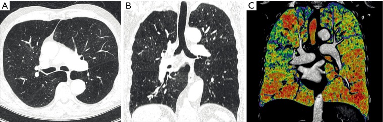
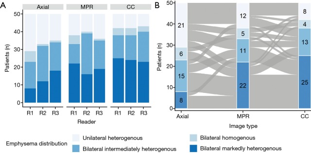
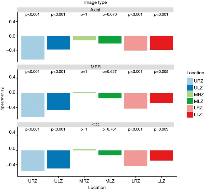
Methods: Fifty patients undergoing lung volume reduction surgery at our institution between September 2015 and February 2016 were retrospectively investigated. Three readers visually assessed the amount and distribution patterns of pulmonary emphysema on axial, multi-planar and CC CT images using the Goddard scoring system and a surgically oriented grading system (bilateral markedly heterogenous, bilateral intermediately heterogenous, bilateral homogenous and unilateral heterogenous emphysema). Observer dependency was investigated by using Fleiss' kappa (κ) and the intraclass correlation coefficient (ICC). Results were compared to quantitative results from densitometry measurements and lung perfusion scintigraphy by using Spearman correlation. Recommendations for lung volume reduction sites based on emphysema amount and distribution of all readers were compared to removal sites from the surgical reports.
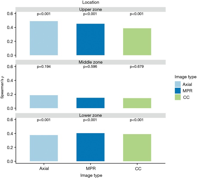
Results: Inter-rater agreement for emphysema distribution rating was substantial for CC images (κ=0.70; 95% CI, 0.64-0.80) and significantly better compared to axial and multiplanar images (P≤0.001). The inter-rater agreement for recommended segment removal was moderate for CC images (κ=0.56; 95% CI, 0.49-0.63) and significantly better compared to axial and multiplanar images (P<0.001). Visual emphysema rating correlated significantly with measurements from densitometry and perfusion scintigraphy in the upper and lower lung zones in all image types.
Conclusions: CC CT images allow a precise, less observer-dependent evaluation of distribution of pulmonary emphysema and resection recommendation compared to axial and multiplanar CT images and might therefore be useful in lung volume resection surgery planning.
Keywords: Pulmonary emphysema; chronic obstructive; densitometry; pneumonectomy; pulmonary disease; tomography, spiral computed.
Conflict of interest statement
Conflicts of Interest: The authors have no conflicts of interest to declare.
Figures
References
-
- The definition of emphysema. Report of a National Heart, Lung, and Blood Institute, Division of Lung Diseases workshop. Am Rev Respir Dis 1985;132:182-5. - PubMed
LinkOut - more resources
Full Text Sources