

Case Reports
doi: 10.1155/2019/6537437.
eCollection 2019.
Bronchoesophageal Fistula due to Esophageal Tuberculosis
Affiliations
- PMID: 31019816
- PMCID: PMC6451813
- DOI: 10.1155/2019/6537437
Item in Clipboard
Case Reports
Bronchoesophageal Fistula due to Esophageal Tuberculosis
Case Rep Infect Dis.
.
Abstract
This is a case report regarding a patient who presented with 6 months of dysphagia and subsequent 40-pound weight loss. The patient underwent imaging, suggestive of pulmonary TB. Further workup of his dysphagia with esophagogastroduodenoscopy and bronchoscopy revealed two bronchoesophageal fistulas. Tuberculosis is an important differential diagnosis of prolonged dysphagia in immunocompetent patients.
Figures
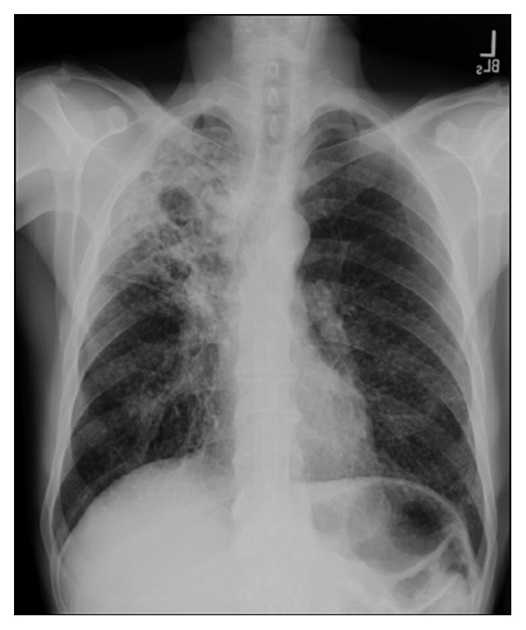
Chest X-ray showing cavitary opacity in the right upper lung field.
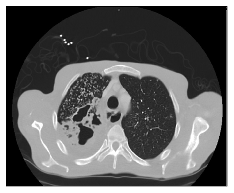
Contrast-enhanced CT scan. Axial section (neck) showing airspace consolidations with diffuse tree in bud opacities in the right lung apex and to a less extent, in the left lung apex. In addition, there are multiple areas of cavities within the right lung apex.
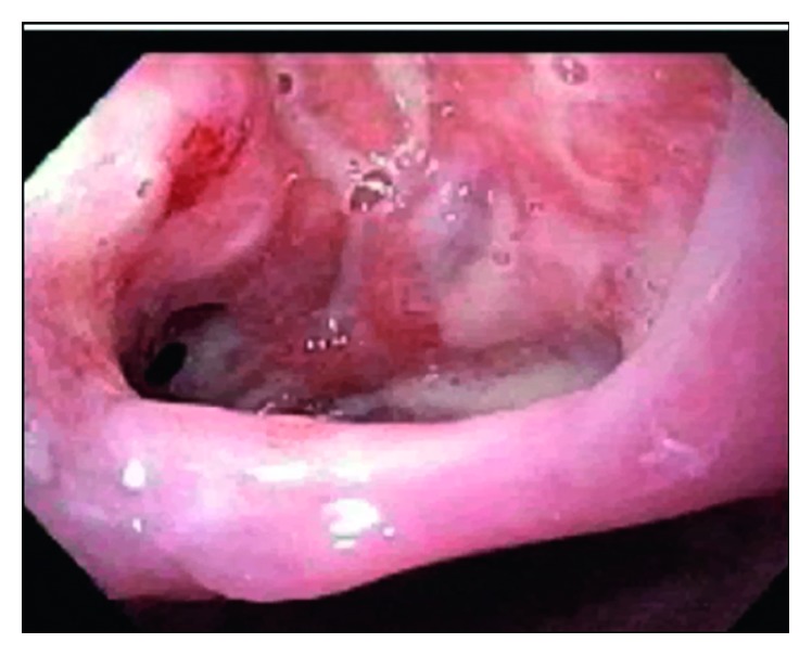
Esophagogastroduodenoscopy (EGD). The upper esophagus at 25 cm showed a 2.5 cm defect with a visible fistulous track to the trachea.
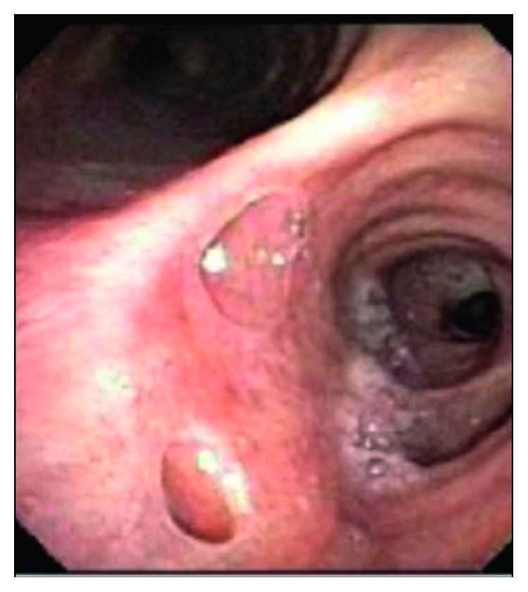
Bronchoscopy. Bronchoesophageal fistulas with openings seen in the right mainstem bronchus on the posterior aspect right below the carina at around 6'o clock position.
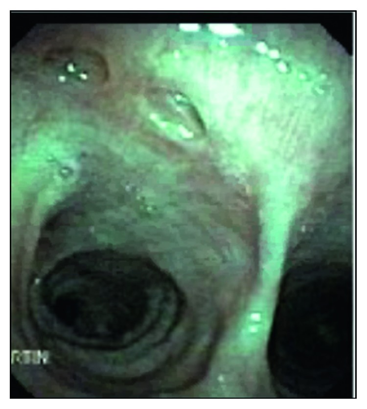
Bronchoscopy (posttreatment). There are two healed fistulas in the right posterior mainstem (posterior to carina), measuring approximately 0.5 cm each.
References
Publication types
LinkOut - more resources
Full Text Sources
