. 2019 Feb 11;8(3):e259-e266.
doi: 10.1016/j.eats.2018.10.022. eCollection 2019 Mar.

Matrix-Induced Autologous Chondrocyte Implantation With Autologous Bone Grafting for Osteochondral Lesions of the Femoral Trochlea

Affiliations

Matrix-Induced Autologous Chondrocyte Implantation With Autologous Bone Grafting for Osteochondral Lesions of the Femoral Trochlea

Kristofer J Jones et al. Arthrosc Tech. .

Abstract

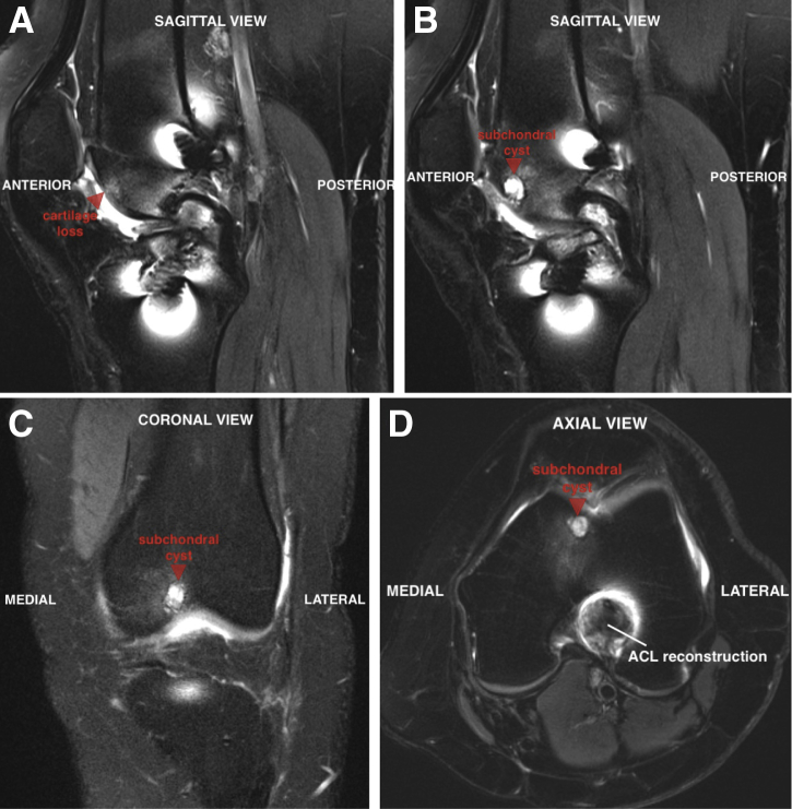
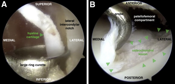
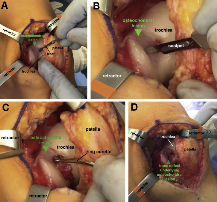
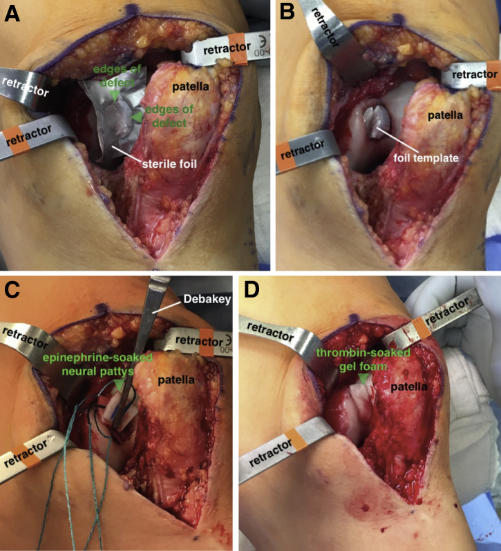
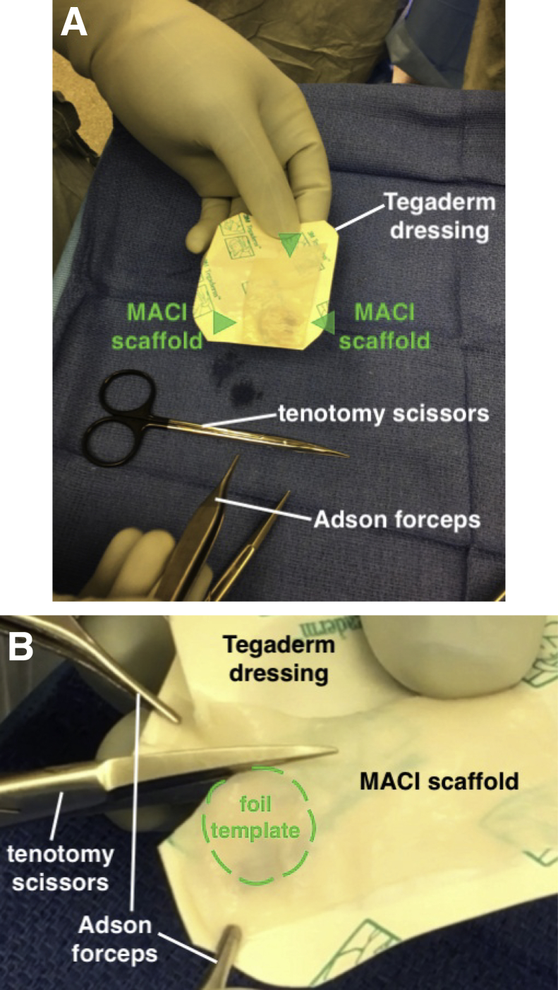
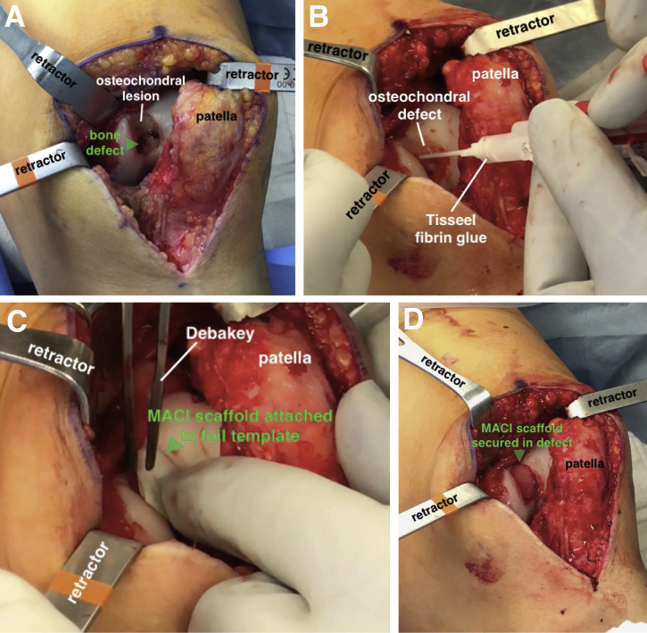
Matrix-induced autologous chondrocyte implantation is a 2-stage surgical procedure used to treat symptomatic, full-thickness chondral lesions of the knee. This third-generation autologous chondrocyte implantation (ACI) technique improves on the limitations of previous methods, including the risk of uneven chondrocyte distribution at the time of implantation and graft hypertrophy. Given the compliant properties of the scaffold, the graft can be easily shaped to treat irregular chondral defects and applied to articular surfaces with multiplanar geometry (e.g., patella, trochlea). Although ACI techniques are ideally suited to treat chondral surface defects, the ACI "sandwich" technique can be used to treat large osteochondral defects with significant bone loss (>8 mm). Historically, this procedure uses autologous bone graft to replace areas of osseous deficiency along with 2 type I/III collagen bilayer membranes to securely contain the cultured chondrocytes within the defect. We present an analogous technique for the treatment of osteochondral lesions of the femoral trochlea using a single matrix-induced ACI scaffold and autologous bone grafting for a segmental osseous defect.


Figures

Fig 1
Fig 1
(A) Preoperative magnetic resonance imaging of the left knee reveals a 1.5-cm region of full-thickness cartilage loss in the femoral trochlea (arrow) on the sagittal view. Underlying the lesion is a 6 × 8-mm subchondral cyst (arrow) that can be seen on sagittal (B), coronal (C), and axial (D) views.
Fig 2
Fig 2
(A) During the first of this 2-stage procedure, a large ring curette is used to harvest 200 to 300 mg of hyaline cartilage (arrow) from the intercondylar notch along the lateral femoral condyle of the left knee with the knee flexed. An anterolateral portal is used for viewing, and the curette is inserted through the anteromedial portal. (B) A diagnostic arthroscopy is also performed to assess the trochlear lesion (arrows) with regard to size, depth, and morphology. This view is from an anterolateral portal with the knee extended.
Fig 3
Fig 3
(A) With the patient supine and left knee slightly flexed, the full-thickness lesion in the femoral trochlea is exposed (arrow) through a medial parapatellar arthrotomy. (B) The borders of the lesion (arrow) are sharply delineated using a 15-blade scalpel. (C) A ring curette is used to debride the base of the defect and create stable vertical edges at the periphery of the lesion (arrow). It is important to remove the underlying calcified cartilage layer without penetrating the subchondral bone. (D) A 2.7-mm drill is used to perforate the lesion and gain access to the underlying bone defect (arrow). The sclerotic margins of the defect are debrided with a curette and drilled with a k-wire.
Fig 4
Fig 4
(A) With the patient supine and left knee slightly flexed, a template is made by placing a sterile foil wrapper from any commercial suture package into the defect and pressing along the edges of the defect (arrows) with a Freer elevator. (B) Once cut, the template can be checked for accurate sizing in the defect, with the 12 o'clock position marked for orientation. After the tourniquet is released, hemostasis at the defect site is achieved with epinephrine-soaked neural patties (C) or thrombin-soaked gel foam (D) (arrows).
Fig 5
Fig 5
(A) The MACI scaffold (arrows) is placed on a Tegaderm dressing to provide a stable platform for graft preparation. When the notch is in the lower left corner of the scaffold, the cellular side is facing up. It is imperative to note proper orientation before cutting the graft. (B) The template is placed between the Tegaderm and the scaffold, and the scaffold is cut using tenotomy scissors. It is helpful to have an assistant hold the corners of the graft and Tegaderm stable with small forceps while cutting. (MACI, matrix-induced autologous chondrocyte implantation.)
Fig 6
Fig 6
(A) With the patient supine and left knee slightly flexed, the defect is filled with BMAC-soaked bone graft to a level flush with the subchondral bone (arrow). (B) The base of the defect is filled with Tisseel fibrin glue. (C) The scaffold is placed flush into the lesion with the cell side down. It is helpful to insert the scaffold with it still attached to the foil template (arrow), to allow for stable insertion in the proper orientation. (D) After removing the template and holding light digital pressure on the graft for 3 minutes, the fibrin glue has solidified and the graft (arrow) is now secured. (BMAC, bone marrow aspirate concentrate.)

Similar articles

Cited by

References

    1. McCormick F., Harris J.D., Abrams G.D. Trends in the surgical treatment of articular cartilage lesions in the United States: An analysis of a large private-payer database over a period of 8 years. Arthroscopy. 2014;30:222–226. - PubMed
    1. Krill M., Early N., Everhart J.S., Flanigan D.C. Autologous chondrocyte implantation (ACI) for knee cartilage defects: A review of indications, technique, and outcomes. JBJS Rev. 2018;6:e5. - PubMed
    1. Brittberg M., Recker D., Ilgenfritz J., Saris D.B.F., SUMMIT Extension Study Group Matrix-applied characterized autologous cultured chondrocytes versus microfracture: Five-year follow-up of a prospective randomized trial. Am J Sports Med. 2018;46:1343–1351. - PubMed
    1. Kreuz P.C., Kalkreuth R.H., Niemeyer P., Uhl M., Erggelet C. Long-term clinical and MRI results of matrix-associated autologous chondrocyte implantation for articular cartilage defects of the knee. Cartilage. February 1, 2018 [E-pub ahead of print.] - PMC - PubMed
    1. Marlovits S., Aldrian S., Wondrasch B. Clinical and radiological outcomes 5 years after matrix-induced autologous chondrocyte implantation in patients with symptomatic, traumatic chondral defects. Am J Sports Med. 2012;40:2273–2280. - PubMed