Case Reports
. 2019 Mar;7(5):108.
doi: 10.21037/atm.2019.02.11.

A case of recurrent hemoptysis caused by pulmonary actinomycosis diagnosed using transbronchial lung biopsy after bronchial artery embolism and a brief review of the literature

Affiliations
Case Reports

A case of recurrent hemoptysis caused by pulmonary actinomycosis diagnosed using transbronchial lung biopsy after bronchial artery embolism and a brief review of the literature

Manabu Suzuki et al. Ann Transl Med. 2019 Mar.

Abstract

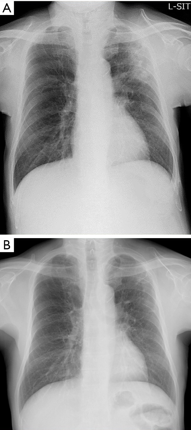
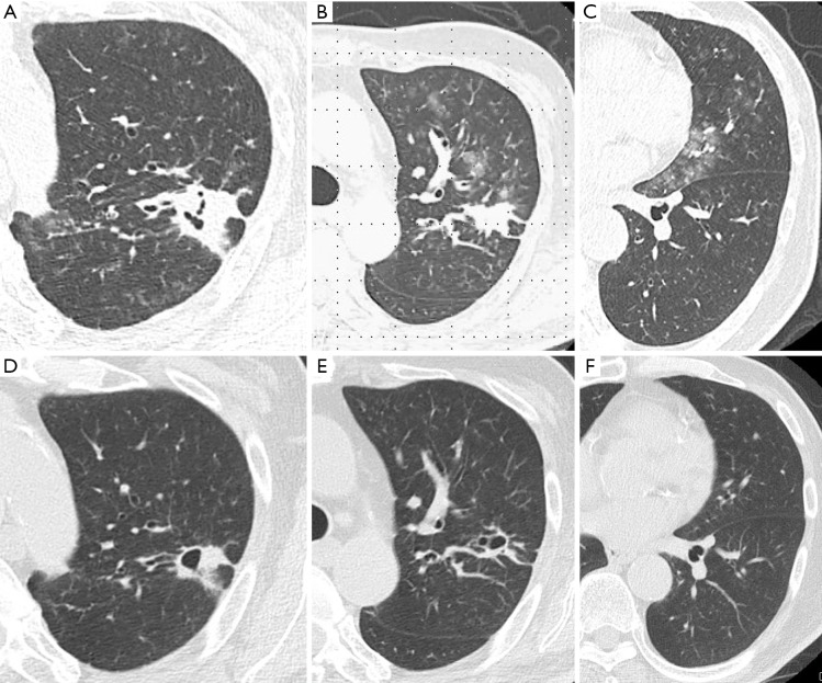
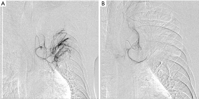
A 60-year-old man was admitted to our hospital because of massive hemoptysis with acute respiratory failure. Since six months ago, he noticed gradual worsening of hemoptysis and was transferred to our hospital. Chest computed tomography showed a nodular lesion with cavitation in the left upper lobe and surrounding ground-glass opacification. Initially, a hemostatic agent was administered, but we eventually performed bronchial artery embolization (BAE) by ourselves due to persistent hemoptysis. After achieving good hemostasis with BAE bronchoscopy was performed, which gave a diagnosis of pulmonary actinomycosis on histopathologic examination of the transbronchial biopsy specimen without the need for lung resection.

Keywords: Recurrent hemoptysis; bronchial artery embolization (BAE); pulmonary actinomycosis; transbronchial lung biopsy.


Conflict of interest statement

Conflicts of Interest: A summary of this paper was presented at the 630th Japan Internal Medical Association Kanto Regional Association (February 2017) and received an encouragement prize.

Figures

Figure 1
Figure 1
Chest radiograph on admission shows widespread infiltration with cavitation in the left upper lobe (A). Chest radiograph after the standard treatment shows widespread that infiltration with cavitation was improved (B).
Figure 2
Figure 2
Chest computed tomography scan reveals a patchy infiltration with cavitation (A,B) in left the upper lobe and surrounding ground glass opacities (C) from hemoptysis. After the completion of the standard treatment, CT scan reveals improved patchy infiltration but remained a cavitation in left the upper lobe (D,E). Ground glass opacities (F) from hemoptysis were improved because of disappeared hemoptysis.
Figure 3
Figure 3
Angiography showing the left bronchial artery. (A) The proximal vessel is dilated and tortuous, with peripheral vascularization and presence of pulmonary artery shunt; (B) after bronchial artery embolization with gelatin sponge particles, the peripheral vascularization and pulmonary artery shunt resolved.
Figure 4
Figure 4
Histopathologic findings. (A) Hematoxylin and eosin stain (4×) revealed dense infiltration of neutrophils, plasma cells, and lymphocytes. (B) Gram stain (40×) revealed Gram-positive bacteria aggregates of a filamentous bacterium, (C) Grocott’s stain (100×) revealed a filamentous bacterium.

References

    1. Smego RA, Jr, Foglia G. Actinomycosis. Clin Infect Dis 1998;26:1255-61. 10.1086/516337 - DOI - PubMed
    1. Valour F, Sénéchal A, Dupieux C, et al. Actinomycosis: etiology, clinical features, diagnosis, treatment, and management. Infect Drug Resist 2014;7:183-97. - PMC - PubMed
    1. Lu MS, Liu HP, Yeh CH, et al. The role of surgery in hemoptysis caused by thoracic actinomycosis; a forgotten disease. Eur J Cardiothorac Surg 2003;24:694-98. 10.1016/S1010-7940(03)00515-3 - DOI - PubMed
    1. Brook I. Actinomycosis: diagnosis and management. South Med J 2008;101:1019-23. 10.1097/SMJ.0b013e3181864c1f - DOI - PubMed
    1. Sakon M, Mikami K, Seki H, et al. A case report of pulmonary actinomycosis radiologically mimicking lung metastasis from rectal cancer. Nihon Rinsho Geka Gakkai Zasshi 2008;69:38-43. 10.3919/jjsa.69.38 - DOI

Publication types