. 2019 Mar 3;6(4):655-668.
doi: 10.1002/acn3.735. eCollection 2019 Apr.

Structural brain anomalies in patients with FOXG1 syndrome and in Foxg1+/- mice

Affiliations

Structural brain anomalies in patients with FOXG1 syndrome and in Foxg1+/- mice

Milka Pringsheim et al. Ann Clin Transl Neurol. .

Abstract

Objective: FOXG1 syndrome is a rare neurodevelopmental disorder associated with heterozygous FOXG1 variants or chromosomal microaberrations in 14q12. The study aimed at assessing the scope of structural cerebral anomalies revealed by neuroimaging to delineate the genotype and neuroimaging phenotype associations.

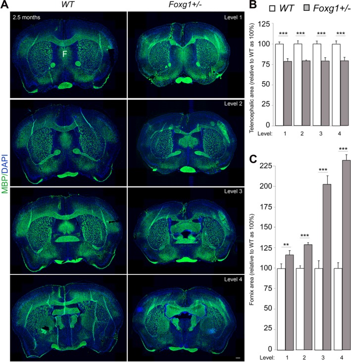
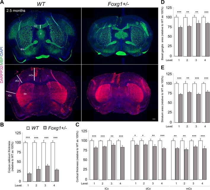
Methods: We compiled 34 patients with a heterozygous (likely) pathogenic FOXG1 variant. Qualitative assessment of cerebral anomalies was performed by standardized re-analysis of all 34 MRI data sets. Statistical analysis of genetic, clinical and neuroimaging data were performed. We quantified clinical and neuroimaging phenotypes using severity scores. Telencephalic phenotypes of adult Foxg1+/- mice were examined using immunohistological stainings followed by quantitative evaluation of structural anomalies.

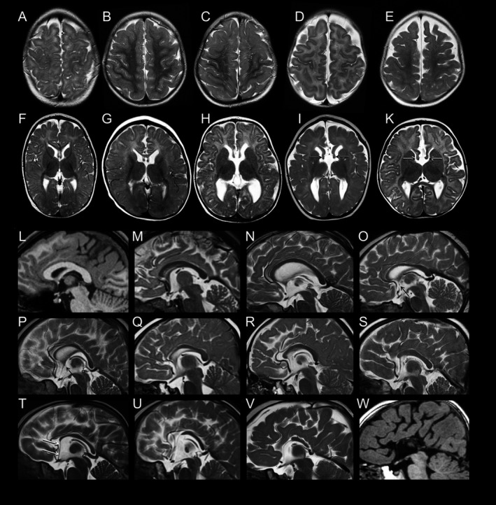
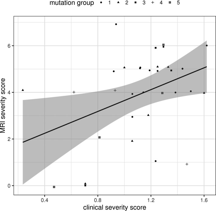
Results: Characteristic neuroimaging features included corpus callosum anomalies (82%), thickening of the fornix (74%), simplified gyral pattern (56%), enlargement of inner CSF spaces (44%), hypoplasia of basal ganglia (38%), and hypoplasia of frontal lobes (29%). We observed a marked, filiform thinning of the rostrum as recurrent highly typical pattern of corpus callosum anomaly in combination with distinct thickening of the fornix as a characteristic feature. Thickening of the fornices was not reported previously in FOXG1 syndrome. Simplified gyral pattern occurred significantly more frequently in patients with early truncating variants. Higher clinical severity scores were significantly associated with higher neuroimaging severity scores. Modeling of Foxg1 heterozygosity in mouse brain recapitulated the associated abnormal cerebral morphology phenotypes, including the striking enlargement of the fornix.

Interpretation: Combination of specific corpus callosum anomalies with simplified gyral pattern and hyperplasia of the fornices is highly characteristic for FOXG1 syndrome.


Conflict of interest statement

Nothing to report.

Figures

Figure 1
Figure 1
Spectrum of structural brain anomalies in FOXG1 syndrome revealed by neuroimaging. Gyral pattern: (A–E) T2‐weighted axial MR images of the fronto‐parietal area in five patients with FOXG1 point mutations show (A, patient #17) normal gyral pattern; (B, #28) mild and (C, #32) moderate simplified gyral pattern; as well as (D, #29; E, #24) simplified gyral pattern with dilated subarachnoid CSF spaces. Basal ganglia and fornices: (F–K) T2‐weighted axial MR images at the level of the basal ganglia show (F, #17) normal basal ganglia, (G, #6) small basal ganglia relative to thalamus, (H, #24; I, #34) dilated ventricles, and (I, #34; K, #8) thickened fornices (arrows). Corpus callosum: (L, W) T1‐weighted and (M through V) T2‐weighted midsagittal MR images show the spectrum of anomalies of the corpus callosum (CC), ranging from (L, #22) normal CC over various degrees (M, #2; N, #33; O, #17; P, #15; Q, #6) of relative thinning of the anterior portions, (R, #18; S, #16) absent rostrum, (T, #8; U, #9; V, #34) partial agenesis of anterior parts to (W, #1) almost complete agenesis. Note the characteristic elongation of the lamina terminalis stretching from the anterior end of the malformed CC to the bottom of the third ventricle (white arrows in T). An anterior commissure is visible only in the milder variants (L–P; black arrows in O, P).
Figure 2
Figure 2
Heterozygous deletion of Foxg1 causes abnormal brain morphology in mice. (A) Immunostaining of MBP and DAPI in coronal sections at different levels (rostral‐caudal) of wild type (WT) and mutant (Foxg1+/−) adult (2.5 months) mouse brain to visualize cross‐section of the entire forebrain and white matter commissural structures like the corpus callosum and fornix (F). (B, C) Bar charts depicting summary of the quantitative analysis of the area of the entire adult mouse telencephalon and fornix, respectively. The cerebral area is significantly reduced (B) while the fornix is conspicuously expanded (C) in mutant brains as compared with controls. The fornix is consistently expanded across its entire structure from rostral to caudal (level 1‐4) (C). Values are presented as means ± SEMs (**P < 0.01, ***P < 0.005). Experimental replicates (n) = 4; Scale bar: 100 μm.
Figure 3
Figure 3
Further discrete cerebral anomalies in Foxg1+/− mouse brain sections. (A) Immunostaining of MBP, DARPP32 and DAPI in coronal sections of wild type (WT) and mutant (Foxg1+/−) adult (2.5 months) mouse brain to visualize gray matter areas such as the Cortex (Cx) and basal ganglia (BG) or Striatum (Str), and white matter commissural systems like the corpus callosum (CC) and fornix (F). (B–E) Bar charts showing summary of the quantitative analysis of forebrain structure alterations in mutants (Foxg1+/−) at various section levels (1‐4) as compared with controls. The thickness of the corpus callosum (B) and distinct cortical domains like the medial cortex (mCx) dorsal cortex (dCx), and lateral cortex (lCx) (C) are significantly reduced in mutants. The areas of the BG (D) and Str (E) are significantly reduced in mutant telencephalon as compared with that of controls. Values are presented as means ± SEMs (*P < 0.05, **P < 0.01, ***P < 0.005). Experimental replicates (n) = 4; Scale bar: 100 μm.
Figure 4
Figure 4
Correlation between clinical severity score and neuroimaging severity score. At the p < 0.05 level there was a significant association found between clinical severity score and neuroimaging severity score (Kendall's rank correlation tau: τ  =  0.27; P = 0.03).

References

    1. Ariani F, Hayek G, Rondinella D, et al. FOXG1 is responsible for the congenital variant of Rett syndrome. Am J Hum Genet 2008;83(1):89–93. - PMC - PubMed
    1. Mencarelli MA, Kleefstra T, Katzaki E, et al. 14q12 Microdeletion syndrome and congenital variant of Rett syndrome. Eur J Med Genet 2009;52:148–152. - PubMed
    1. Kortüm F, Das S, Flindt M, et al. The core FOXG1 syndrome phenotype consists of postnatal microcephaly, severe mental retardation, absent language, dyskinesia, and corpus callosum hypogenesis. J Med Genet 2011;48:396–406. - PMC - PubMed
    1. Philippe C, Amsallem D, Francannet C, et al. Phenotypic variability in Rett syndrome associated with FOXG1 mutations in females. J Med Genet 2010;47:59–65. - PubMed
    1. De Filippis R, Pancrazi L, Bjørgo K, et al. Expanding the phenotype associated with FOXG1 mutations and in vivo FoxG1 chromatin‐binding dynamics. Clin Genet 2012;82(4):395–403. - PubMed

LinkOut - more resources