. 2019 Apr;7(8):e14056.
doi: 10.14814/phy2.14056.

Peptone-mediated glucagon-like peptide-1 secretion depends on intestinal absorption and activation of basolaterally located Calcium-Sensing Receptors

Affiliations

Peptone-mediated glucagon-like peptide-1 secretion depends on intestinal absorption and activation of basolaterally located Calcium-Sensing Receptors

Ida M Modvig et al. Physiol Rep. 2019 Apr.

Abstract

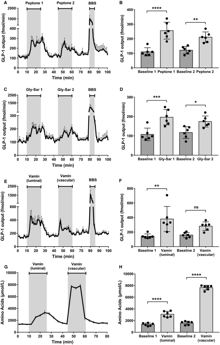
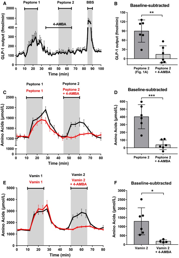
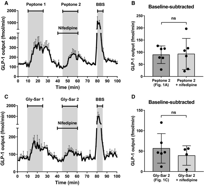
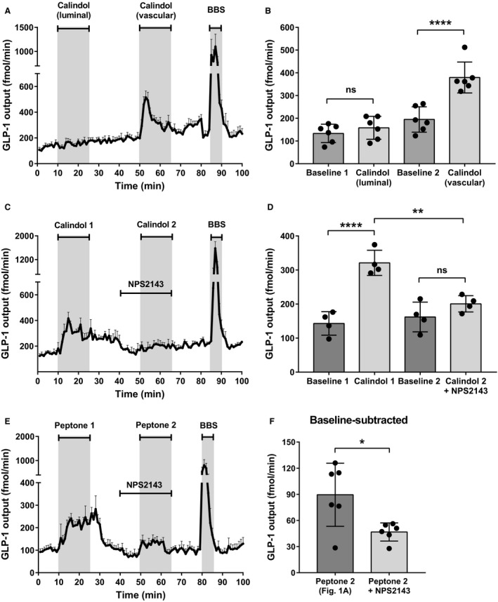
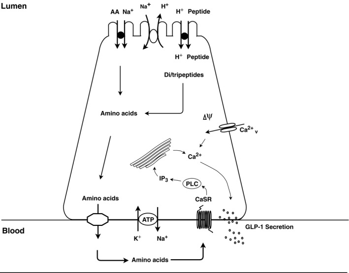
Protein intake robustly stimulates the secretion of the incretin hormone, glucagon-like peptide-1 (GLP-1) but the molecular mechanisms involved are not well understood. In particular, it is unknown whether proteins stimulate secretion by activation of luminal or basolateral sensors. We characterized the mechanisms using a physiologically relevant model - the isolated perfused proximal rat small intestine. Intraluminal protein hydrolysates derived from meat (peptone; 50 mg/mL) increased GLP-1 secretion 2.3-fold (from a basal secretion of 110 ± 28 fmol/min). The sensory mechanisms underlying the response depended on di/tripeptide uptake through Peptide Transporter 1 (PepT1) and subsequent basolateral activation of the amino acid sensing receptor, Calcium-Sensing Receptor (CaSR), since inhibition of PepT1 as well as CaSR both attenuated the peptone-induced GLP-1 response. Supporting this, intraluminal peptones were absorbed efficiently by the perfused intestine (resulting in increased amino acid concentrations in the venous effluent) and infusion of amino acids robustly stimulated GLP-1 secretion. Inhibitors of voltage-gated L-type Ca2+ channels had no effect on secretion suggesting that peptone-mediated GLP-1 secretion is not mediated by L-cell depolarization with subsequent opening of these channels. Specific targeting of CaSR could serve as a target to stimulate the endogenous secretion of GLP-1.

Keywords: Amino acid sensing; Calcium-Sensing Receptor; glucagon-like peptide 1; peptide transporter 1; peptone.


Conflict of interest statement

The authors of this work declare no potential conflicts of interest relevant to this article.

Figures

Figure 1
Figure 1
Luminal peptone stimulates GLP‐1 secretion from the proximal small intestine. (A) Total GLP‐1 outputs shown as means ± SEM. Peptone (50 mg/mL) was infused intraluminally between minute 11–25 and minute 46–60. Bombesin (BBS; 10 nmol/L), a positive control, was infused between minute 80–85. (B) Mean GLP‐1 outputs at baseline (Baseline 1 and 2) and following stimulation with luminal peptones (Peptone 1 and 2). (C) Total GLP‐1 outputs shown as means ± SEM. Gly‐Sar (22 mg/mL) was infused intraluminally between minute 11–25 and minute 46–60 (D) Mean GLP‐1 outputs at baseline (baseline 1 and 2) and following stimulation with luminal Gly‐Sar (Gly‐Sar 1 and 2). (E) Total GLP‐1 outputs shown as means ± SEM. Vamin (51 mg/mL) was infused intraluminally between minute 11–25. Vamin (4.25 mg/mL) was infused intravascularly between minute 46–60. (F) Mean GLP‐1 outputs at baseline (baseline 1 and 2) and following stimulation with luminal and vascular Vamin, respectively. (G) Total amino acid concentrations in venous effluent shown as means ± SEM. (H) Mean concentrations of total amino acids at baseline (baseline 1 and baseline 2) and following stimulation with, respectively, luminally and vascularly administered Vamin. Statistical significance was tested by one‐way ANOVA for repeated measurements followed by Bonferroni post hoc test; NS: nonsignificant: P > 0.05. *P < 0.05, **P < 0.01, ***P < 0.001, ****P < 0.0001. n = 6 for all groups.
Figure 2
Figure 2
Blockage of PepT1 inhibits peptone‐induced GLP‐1 secretion and peptide absorption. (A) Total GLP‐1 outputs shown as means ± SEM. Peptone (50 mg/mL) was intraluminally infused between minute 11–25 and minute 51–65. A PepT1 blocker, 4‐AMBA (20 mmol/L), was intraluminally infused between min 36–65. (B) Mean baseline‐subtracted GLP‐1 outputs in response to peptone stimulation with and without 4‐AMBA (Peptone 2; Fig. 1 vs. Peptone 2 + 4‐AMBA). (C) Total amino acid concentrations in venous effluent shown as means ± SEM. Peptone (50 mg/mL) was intraluminally infused between minute 11–25 and minute 46–65. A PepT1 blocker, 4‐AMBA (20 mmol/L), was intraluminally infused between min 36–65. (D) Mean amino acid concentrations subtracted baseline during peptone stimulation with and without 4‐AMBA (Peptone 2 vs. Peptone 2 + 4‐AMBA). (E) Total amino acid concentrations in venous effluent shown as means ± SEM. Vamin (51 mg/mL) was intraluminally infused between minute 11–25 and minute 46–65. A PepT1 blocker, 4‐AMBA (20 mmol/L), was intraluminally infused between min 36–65. (F) Mean amino acid concentrations (after subtraction of baseline values) during Vamin stimulation with and without 4‐AMBA (Vamin 2 vs. Vamin 2 + 4‐AMBA). Statistical significance was tested by an unpaired student's t‐test; NS: nonsignificant: P > 0.05. *P < 0.05, **P < 0.01, ***P < 0.001, ****P < 0.0001. n = 6 for all groups except Vamin + 4‐AMBA (Fig. 2E), n = 4.
Figure 3
Figure 3
L‐type Ca2+‐channels are not involved in peptone‐stimulated GLP‐1 secretion. (A) Total GLP‐1 outputs shown as means ± SEM. Peptone (50 mg/mL) was intraluminally infused between minute 11–25 and minute 46–60. Nifedipine (10 μmol/L), an L‐type Cav 2+‐channel inhibitor, was infused intravascularly between minute 40–60. (B) Mean values of the baseline‐subtracted peptone‐stimulated GLP‐1 outputs with and without nifedipine (Peptone 2; Fig. 1 vs. Peptone 2 + nifedipine). (C) Total GLP‐1 outputs shown as means ± SEM. Gly‐Sar (22 mg/mL) was intraluminally infused between minute 11–25 and minute 46–60. Nifedipine (10 μmol/L) was infused intravascularly between minute 40–60. (D) Mean values of the baseline‐subtracted Gly‐Sar‐stimulated GLP‐1 outputs with and without nifedipine (Gly‐Sar 2; Fig. 1 vs. Gly‐Sar 2 + nifedipine). Statistical significance was tested by an unpaired student's t‐test; NS: nonsignificant: P > 0.05. *P < 0.05, **P < 0.01, ***P < 0.001, ****P < 0.0001. n = 6 for all groups except Gly‐Sar + nifedipine, n = 4.
Figure 4
Figure 4
Calcium‐Sensing Receptor is located on the basolateral membrane and is involved in peptone‐mediated GLP‐1 secretion. (A) Total GLP‐1 outputs shown as means ±  SEM. Calindol (50 μmol/L), a positive allosteric modulator of CaSR, was intraluminally infused between minute 11–25 and intravascularly infused between minute 51–65. (B) Mean GLP‐1 outputs at baseline (Baseline 1 and 2) and following stimulation with luminal calindol and vascular calindol. (C) Total GLP‐1 outputs shown as means ± SEM. Calindol (50 μmol/L) was intraluminally infused between minute 11–25 and intravascular infused between minute 51–65. NPS2143 (25 μmol/L), a negative allosteric modulator of CaSR, was intravascularly infused between minute 41–60. (D) Mean GLP‐1 outputs at baseline (Baseline 1 and 2) and following stimulation with vascular calindol (Calindol 1) and calindol + NPS2143 (Calindol 2 + NPS2143). (E) Total GLP‐1 outputs shown as means ± SEM. Peptone (50 mg/mL) was intraluminally infused between minute 11–25 and 51–65. NPS2143 (25 μmol/L), was intravascularly infused between minute 41–60. (F) Mean values of baseline‐subtracted peptone‐stimulated GLP‐1 secretion with and without NPS2143 (Peptone 2; Fig. 1 vs. Peptone 2 + NPS2143). Statistical significance was tested by one‐way ANOVA for repeated measurements followed by Bonferroni post hoc test (Fig. 4B+D) and by unpaired student's t‐test (Fig. 4F); NS: nonsignificant: P > 0.05. *P < 0.05, **P < 0.01, ***P < 0.001, ****P < 0.0001. n = 6 for all groups except Calindol + NPS2143 (Fig. 4C), n = 4.
Figure 5
Figure 5
Illustration of the endocrine L cell and the proposed mechanisms by which peptone stimulates GLP‐1 release. Di/tripeptides are taken up by PepT1 and are degraded by cytosolic peptidases to their respective amino acids. In addition, free amino acids present in the intestinal lumen may be taken up by different amino acid transporters. Intracellular amino acids are then transported to the interstitial side through basolateral amino acid transporters, wherefrom they stimulate the L cells by activating amino acid sensors, like CaSR, situated on the basolateral membrane.

References

    1. Alamshah, A. , Spreckley E., Norton M., Kinsey‐Jones J. S., Amin A., Ramgulam A., et al. 2017. l‐phenylalanine modulates gut hormone release and glucose tolerance, and suppresses food intake through the calcium‐sensing receptor in rodents. Int. J. Obes. 41:1693–1701. - PMC - PubMed
    1. Baggio, L. L. , and Drucker D. J.. 2007. Biology of incretins: GLP‐1 and GIP. Gastroenterology 132:2131–2157. - PubMed
    1. Belza, A. , Ritz C., Sørensen M. Q., Holst J. J., Rehfeld J. F., and Astrup A.. 2013. Contribution of gastroenteropancreatic appetite hormones to protein‐induced satiety. Am. J. Clin. Nutr. 97:980–989. - PubMed
    1. Clemmensen, C. , Jørgensen C. V., Smajilovic S., and Bräuner‐Osborne H.. 2017. Robust GLP‐1 secretion by basic L‐amino acids does not require the GPRC6A receptor. Diabetes Obes. Metab. 19:599–603. - PubMed
    1. Conigrave, A. D. , and Hampson D. R.. 2006. Broad‐spectrum l‐amino acid sensing by class 3 G‐protein‐coupled receptors. Trends Endocrinol. Metab. 17:398–407. - PubMed

Publication types

LinkOut - more resources