. 2019 Aug;10(4):903-918.
doi: 10.1002/jcsm.12434. Epub 2019 Apr 24.

Amelioration of muscle wasting by glucagon-like peptide-1 receptor agonist in muscle atrophy

Affiliations

Amelioration of muscle wasting by glucagon-like peptide-1 receptor agonist in muscle atrophy

Yeonhee Hong et al. J Cachexia Sarcopenia Muscle. 2019 Aug.

Abstract

Background: Skeletal muscle atrophy is defined as a reduction of muscle mass caused by excessive protein degradation. However, the development of therapeutic interventions is still in an early stage. Although glucagon-like peptide-1 receptor (GLP-1R) agonists, such as exendin-4 (Ex-4) and dulaglutide, are widely used for the treatment of diabetes, their effects on muscle pathology are unknown. In this study, we investigated the therapeutic potential of GLP-1R agonist for muscle wasting and the mechanisms involved.

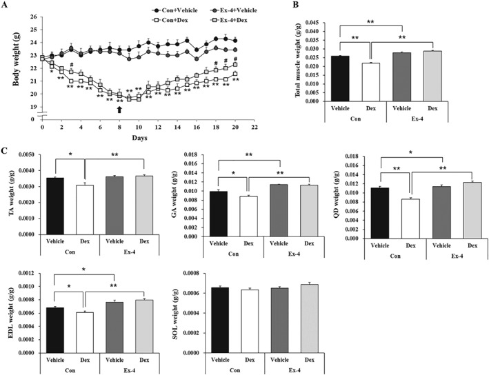
Methods: Mouse C2C12 myotubes were used to evaluate the in vitro effects of Ex-4 in the presence or absence of dexamethasone (Dex) on the regulation of the expression of muscle atrophic factors and the underlying mechanisms using various pharmacological inhibitors. In addition, we investigated the in vivo therapeutic effect of Ex-4 in a Dex-induced mouse muscle atrophy model (20 mg/kg/day i.p.) followed by injection of Ex-4 (100 ng/day i.p.) for 12 days and chronic kidney disease (CKD)-induced muscle atrophy model. Furthermore, we evaluated the effect of a long-acting GLP-1R agonist by treatment of dulaglutide (1 mg/kg/week s.c.) for 3 weeks, in DBA/2J-mdx mice, a Duchenne muscular dystrophy model.

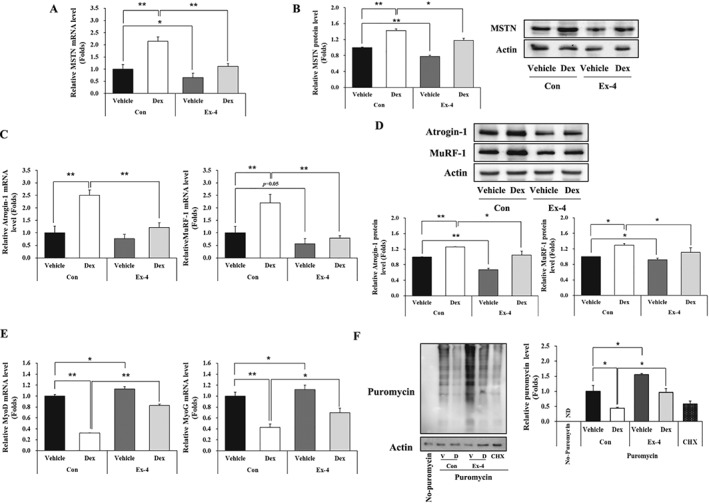
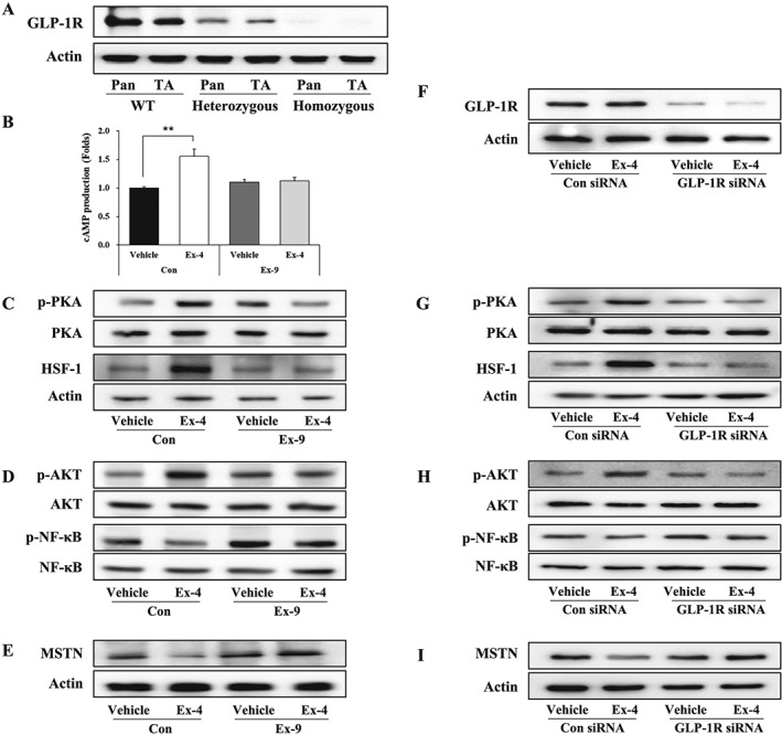
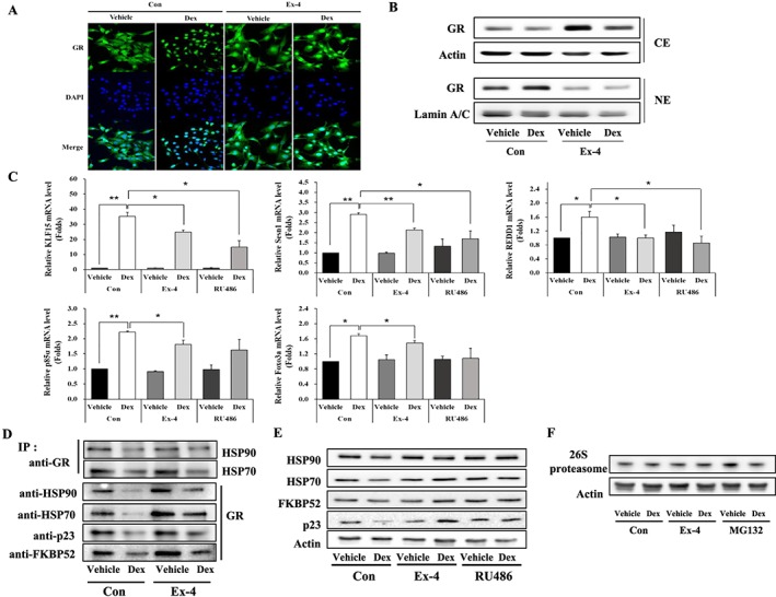
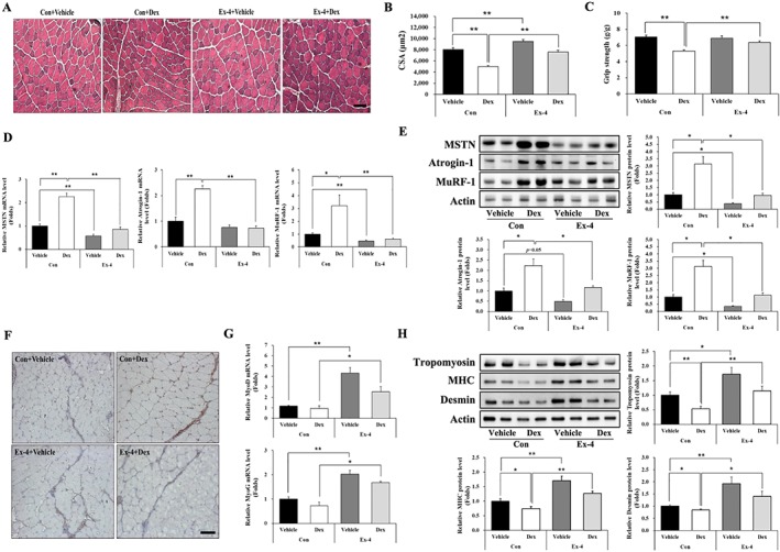
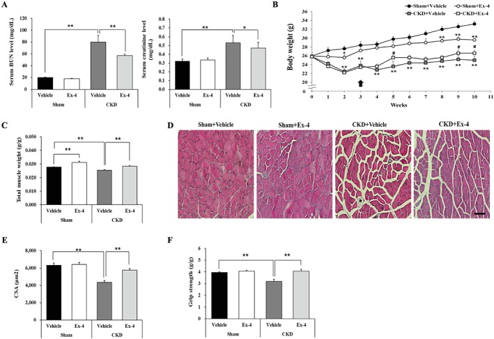
Results: Ex-4 suppressed the expression of myostatin (MSTN) and muscle atrophic factors such as F-box only protein 32 (atrogin-1) and muscle RING-finger protein-1 (MuRF-1) in Dex-treated C2C12 myotubes. The suppression effect was via protein kinase A and protein kinase B signalling pathways through GLP-1R. In addition, Ex-4 treatment inhibited glucocorticoid receptor (GR) translocation by up-regulating the proteins of GR inhibitory complexes. In a Dex-induced muscle atrophy model, Ex-4 ameliorated muscle atrophy by suppressing muscle atrophic factors and enhancing myogenic factors (MyoG and MyoD), leading to increased muscle mass and function. In the CKD muscle atrophy model, Ex-4 also increased muscle mass, myofiber size, and muscle function. In addition, treatment with a long-acting GLP-1R agonist, dulaglutide, recovered muscle mass and function in DBA/2J-mdx mice.

Conclusions: GLP-1R agonists ameliorate muscle wasting by suppressing MSTN and muscle atrophic factors and enhancing myogenic factors through GLP-1R-mediated signalling pathways. These novel findings suggest that activating GLP-1R signalling may be useful for the treatment of atrophy-related muscular diseases.

Keywords: Chronic kidney disease; Dexamethasone; Duchenne muscular dystrophy; GLP-1R agonists; Glucocorticoid receptor; Skeletal muscle atrophy.


Conflict of interest statement

Y.H.H., J.H.L., K.W.J., C.S.C., and H.‐S.J. declare that they have no conflict of interest.

Figures

Figure 1
Figure 1
Ex‐4 suppresses MSTN‐mediated muscle atrophic factors in C2C12 myotubes. C2C12 myotubes differentiated for 5 days were treated with 1 μM Dex at 6 h earlier prior to treatment of with or without 20 nM Ex‐4 for 6 h. (A) The mRNA level of MSTN. (B) The protein level of MSTN with a representative blot. (C) The mRNA level of muscle atrophic factors (atrogin‐1 and MuRF‐1). (D) The protein level of muscle atrophic factors (atrogin‐1 and MuRF‐1) with a representative blot. (E) The mRNA level of myogenic factors (MyoD and MyoG). (F) The puromycin‐labelled protein levels with a representative blot. All values are expressed as the mean ± standard error of the fold‐change relative to controls. Significant differences are indicated as **P < 0.01, *P < 0.05 compared with Con + vehicle or Con + Dex. n = 3. Con, control; Dex, dexamethasone; Ex‐4, exendin‐4; CHX, cycloheximide; ND, not detected.
Figure 2
Figure 2
Ex‐4 regulates the expression of MSTN through GLP‐1R‐mediated PKA and AKT signalling pathways. (A) The protein expression level of GLP‐1R in the pancreas and TA muscle from WT (Glp1r +/+ Cre ), heterozygous (Glp1r flox/+ Cre +), and homozygous (Glp1r flox/flox Cre +) mice with a representative blot. (B) C2C12 myotubes were pretreated with 20 nM of Ex‐9 (GLP‐1R antagonist) for 10 min and then were further treated with 20 nM of Ex‐4 for 30 min. cAMP production was measured using cAMP ELISA kit. (C–E) The treated C2C12 myotubes were examined for the activation of GLP‐1R downstream mediators including PKA and HSF‐1 (C) and AKT and NF‐κB (D) signals using western blot. (E) The protein level of MSTN with a representative blot. (F–I) GLP‐1R siRNA‐transfected C2C12 myotubes were treated with 20 nM Ex‐4 for 30 min. (F) Representative blot of GLP‐1R protein levels. (G–I) C2C12 myotubes were examined for the activation of downstream mediators of GLP‐1R, including PKA and HSF‐1 (G) and AKT and NF‐κB (H) by western blotting. (I) Representative blot of MSTN protein levels. All values are expressed as the mean ± standard error. Significant differences are indicated as **P < 0.01 compared with Con + vehicle. n = 3. Con, control; Ex‐4, exendin‐4; Ex‐9, exendin‐9.
Figure 3
Figure 3
Ex‐4 inhibits the translocation of GR from cytosol into nucleus by up‐regulating the proteins of GR inhibitory complexes. (A) C2C12 myotubes treated with 1 μM Dex at 6 h prior to treatment of with or without 20 nM Ex‐4 for 6 h. GR expression was determined by immunostaining with anti‐GR (green). The nucleus was detected by 4′,6‐diamidino‐2‐phenylindole (DAPI) staining (blue) (magnification ×400). (B) The treated C2C12 myotubes were separated into cytosolic extract (CE) and nuclear extract (NE). The protein level of GR was measured in CE and NE using western blotting. (C) The differentiated C2C12 myotubes treated with 1 μM of Dex at 6 h earlier prior to treatment with 20 nM of either Ex‐4 or RU486, a GR inhibitor, for 6 h. The mRNA level of GR‐targeted genes (KLF15, Sesn1, REDD1, p85α, and FoxO3a) were assessed by RT‐QPCR. (D) The whole cell lysate was isolated and then immunoprecipitated with appropriate antibodies. IP assay of the interaction between GR and GR inhibitory complex proteins (HSP70, HSP90, FKBP52, and p23). (E) The protein level of GR inhibitory complexes in CE. C2C12 myotubes were pretreated with 1 μM Dex and then 6 h later incubated with 20 nM Ex‐4 or RU486 until 12 h. The CE were isolated and then subjected to western blotting and probing with appropriate antibodies. (F) 26S proteasome level was assessed by western blotting. All value expressed as the mean ± standard error. Significant differences are indicated as **P < 0.01, *P < 0.05 compared with Con + vehicle or Con + Dex. n = 3. Con, control; Dex, dexamethasone; Ex‐4, exendin‐4.
Figure 4
Figure 4
Ex‐4 increases muscle mass in Dex‐induced muscle atrophy mice. (A) Body weight was determined during treatment of period. Ten‐week‐old C57BL/6 male mice were administered the Dex (20 mg/kg i.p.) daily for 8 days and then were administered Ex‐4 (100 ng/mouse i.p.) daily for 12 days. Black arrow indicates the start of Ex‐4 injection. Significant differences are indicated as **P < 0.01, *P < 0.05 compared with Con + vehicle; ##P < 0.01, #P < 0.05 compared with Con + Dex. n = 5–8/group. (B) The weight of total muscle tissue. The total muscle weight was normalized to the final body weight (g). (C) The weight of each muscle, including gastrocnemius (GA), tibialis anterior (TA), quadriceps (QD), extensor digitorum longus (EDL), and soleus (SOL), were measured right after sacrifice and normalized to the final body weight (g). All values are expressed as the mean ± standard error. Significant differences are indicated as **P < 0.01, *P < 0.05 compared with Con + vehicle or Con + Dex. n = 5–8/group. Con, control; Dex, dexamethasone; Ex‐4, exendin‐4.
Figure 5
Figure 5
Ex‐4 recovers muscle strength in Dex‐induced muscle atrophy mice. (A) Frozen serial transverse cryosections (7 μm) from TA muscle tissue were stained with H&E and examined under a microscope (magnification ×200). (B) The cross‐sectional area (CSA) of muscle fibre was measured using Image J program. (C) Muscle function was assessed using grip strength measurement. The grip strength was normalized to the final body weight (g). (D–E) The mRNA level of muscle atrophic factors (MSTN, atrogin‐1, and MuRF‐1) (D) and protein (E) levels were assessed using RT‐QPCR and western blotting in TA muscle tissue. (F) The TA muscle tissue from mice administered with Dex was immunostained with anti‐MSTN antibody. The image was taken under a confocal microscope (magnification ×200). Brown colour indicates MSTN expression. (G) The mRNA levels of myogenic factors (MyoD and MyoG) were assessed using RT‐QPCR in TA muscle tissue. (H) The protein levels of myofibrillar proteins (tropomyosin, MHC, and desmin) were measured using western blot. All values are expressed as the mean ± standard error. Significant differences are indicated as **P < 0.01, *P < 0.05 compared with Con + vehicle or Con + Dex, n = 5/group. Con, control; Dex, dexamethasone; Ex‐4, exendin‐4.
Figure 6
Figure 6
Ex‐4 increases total muscle mass and improves muscle functions in CKD mice. Ten‐week‐old C57BL/6 male mice underwent two‐step subtotal nephrectomy to induce muscle atrophy. (A) The serum level of blood urea nitrogen (BUN) and creatinine. (B) The body weight change during treatment. Black arrow indicates the start of Ex‐4 injection. Significant differences are indicated as **P < 0.01, *P < 0.05 compared with sham + vehicle; ##P < 0.01, #P < 0.05 compared with CKD + vehicle. (C) The weight of total muscle tissue. Total muscle weight was normalized to final body weight (g). (D) Frozen serial transverse cryosections (7 μm) of TA muscle were stained with H&E and examined under a microscope (magnification ×200). (E) The CSA of muscle fibre was measured using Image J program. (F) The grip strength was normalized to the final body weight (g). All values are expressed as the mean ± standard error. Significant differences are indicated as **P < 0.01, *P < 0.05 compared with sham + vehicle or CKD + vehicle. n = 5–7/group.
Figure 7
Figure 7
Dulaglutide improves muscle strength in DBA/2J‐mdx mice. Seven‐week‐old DBA/2J‐mdx male mice were subcutaneously administered with 1 mg/kg of dulaglutide once a week for 3 weeks. (A) Sections of TA muscle were stained with H&E and examined under a microscope (magnification ×200). (B) The CSA of muscle fibre was measured using Image J program. (C) The grip strength before administration of dulaglutide. (D) The grip strength after administration of dulaglutide. The grip strength was normalized to the final body weight (g). (E) Four‐limb hanging test after administration of dulaglutide. The left bar graph shows the average of total hanging time of each group; the right dot graph shows the distribution of individual mice for hanging time. All values are expressed as the mean ± standard error. Significant differences are indicated as **P < 0.01, *P < 0.05 compared with vehicle. n = 9–10/group.

References

    1. Cohen S, Nathan JA, Goldberg AL. Muscle wasting in disease: molecular mechanisms and promising therapies. Nat Rev Drug Discov 2015;14:58–74. - PubMed
    1. Workeneh BT, Mitch WE. Review of muscle wasting associated with chronic kidney disease. Am J Clin Nutr 2010;91:1128s–1132s. - PubMed
    1. Evans WJ, Morley JE, Argilés J, Bales C, Baracos V, Guttridge D, et al. Cachexia: a new definition. Clin Nutr 2008;27:793–799. - PubMed
    1. Williams A, Sun X, Fischer JE, Hasselgren PO. The expression of genes in the ubiquitin‐proteasome proteolytic pathway is increased in skeletal muscle from patients with cancer. Surgery 1999;126:744–749, discussion 749‐50, 750. - PubMed
    1. Muscaritoli M, Anker SD, Argiles J, Aversa Z, Bauer JM, Biolo G, et al. Consensus definition of sarcopenia, cachexia and pre‐cachexia: joint document elaborated by Special Interest Groups (SIG) “cachexia‐anorexia in chronic wasting diseases” and “nutrition in geriatrics”. Clin Nutr 2010;29:154–159. - PubMed

Publication types

Substances